5AMH | pdb_00005amh

Cereblon isoform 4 from Magnetospirillum gryphiswaldense in complex with Thalidomide, trigonal crystal form


Experimental Data Snapshot

  • Method: X-RAY DIFFRACTION
  • Resolution: 1.20 Å
  • R-Value Free: 
    0.157 (Depositor), 0.166 (DCC) 
  • R-Value Work: 
    0.126 (Depositor), 0.136 (DCC) 
  • R-Value Observed: 
    0.127 (Depositor) 

Starting Model: experimental
View more details

wwPDB Validation 3D Report Full Report

Validation slider image for 5AMH

Ligand Structure Quality Assessment 


This is version 1.1 of the entry. See complete history

Literature

Structural Dynamics of the Cereblon Ligand Binding Domain.

Hartmann, M.D.Boichenko, I.Coles, M.Lupas, A.N.Hernandez Alvarez, B.

(2015) PLoS One 10: 28342

  • DOI: https://doi.org/10.1371/journal.pone.0128342
  • Primary Citation Related Structures: 
    5AMH, 5AMI, 5AMJ, 5AMK

  • PubMed Abstract: 

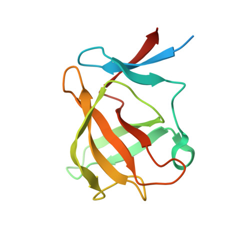
    Cereblon, a primary target of thalidomide and its derivatives, has been characterized structurally from both bacteria and animals. Especially well studied is the thalidomide binding domain, CULT, which shows an invariable structure across different organisms and in complex with different ligands. Here, based on a series of crystal structures of a bacterial representative, we reveal the conformational flexibility and structural dynamics of this domain. In particular, we follow the unfolding of large fractions of the domain upon release of thalidomide in the crystalline state. Our results imply that a third of the domain, including the thalidomide binding pocket, only folds upon ligand binding. We further characterize the structural effect of the C-terminal truncation resulting from the mental-retardation linked R419X nonsense mutation in vitro and offer a mechanistic hypothesis for its irresponsiveness to thalidomide. At 1.2Å resolution, our data provide a view of thalidomide binding at atomic resolution.


  • Organizational Affiliation
    • Department of Protein Evolution, Max Planck Institute for Developmental Biology, Tübingen, Germany.

Macromolecule Content 

  • Total Structure Weight: 14.37 kDa 
  • Atom Count: 1,060 
  • Modeled Residue Count: 106 
  • Deposited Residue Count: 125 
  • Unique protein chains: 1

Macromolecules

Find similar proteins by:|  3D Structure
Entity ID: 1
MoleculeChains  Sequence LengthOrganismDetailsImage
CEREBLON ISOFORM 4125Magnetospirillum gryphiswaldense MSR-1Mutation(s): 0 
UniProt
Find proteins for A4TVL0 (Magnetospirillum gryphiswaldense)
Explore A4TVL0 
Go to UniProtKB:  A4TVL0
Entity Groups
Sequence Clusters30% Identity50% Identity70% Identity90% Identity95% Identity100% Identity
UniProt GroupA4TVL0
Sequence Annotations
Expand
Reference Sequence

Small Molecules

Ligands 5 Unique
IDChains Name / Formula / InChI Key2D Diagram3D Interactions
EF2

Query on EF2


Download:Ideal Coordinates CCD File
C [auth A]S-Thalidomide
C13 H10 N2 O4
UEJJHQNACJXSKW-VIFPVBQESA-N
ZN

Query on ZN


Download:Ideal Coordinates CCD File
B [auth A]ZINC ION
Zn
PTFCDOFLOPIGGS-UHFFFAOYSA-N
NI

Query on NI


Download:Ideal Coordinates CCD File
L [auth A]NICKEL (II) ION
Ni
VEQPNABPJHWNSG-UHFFFAOYSA-N
CA

Query on CA


Download:Ideal Coordinates CCD File
D [auth A]CALCIUM ION
Ca
BHPQYMZQTOCNFJ-UHFFFAOYSA-N
CL

Query on CL


Download:Ideal Coordinates CCD File
E [auth A]
F [auth A]
G [auth A]
H [auth A]
I [auth A]
E [auth A],
F [auth A],
G [auth A],
H [auth A],
I [auth A],
J [auth A],
K [auth A]
CHLORIDE ION
Cl
VEXZGXHMUGYJMC-UHFFFAOYSA-M

Experimental Data & Validation

Experimental Data

  • Method: X-RAY DIFFRACTION
  • Resolution: 1.20 Å
  • R-Value Free:  0.157 (Depositor), 0.166 (DCC) 
  • R-Value Work:  0.126 (Depositor), 0.136 (DCC) 
  • R-Value Observed: 0.127 (Depositor) 
Space Group: P 32 2 1
Unit Cell:
Length ( Å )Angle ( ˚ )
a = 51.72α = 90
b = 51.72β = 90
c = 84.71γ = 120
Software Package:
Software NamePurpose
REFMACrefinement
XDSdata reduction
XDSdata scaling
MOLREPphasing

Structure Validation

View Full Validation Report


Ligand Structure Quality Assessment 


Entry History 

Deposition Data

Revision History  (Full details and data files)

  • Version 1.0: 2015-07-01
    Type: Initial release
  • Version 1.1: 2024-01-10
    Changes: Data collection, Database references, Derived calculations, Other, Refinement description