5AD1 | pdb_00005ad1

A complex of the synthetic siderophore analogue Fe(III)-8-LICAM with the CeuE periplasmic protein from Campylobacter jejuni


Experimental Data Snapshot

  • Method: X-RAY DIFFRACTION
  • Resolution: 1.32 Å
  • R-Value Free: 
    0.181 (Depositor), 0.188 (DCC) 
  • R-Value Work: 
    0.140 (Depositor), 0.152 (DCC) 
  • R-Value Observed: 
    0.142 (Depositor) 

Starting Model: experimental
View more details

wwPDB Validation 3D Report Full Report

Validation slider image for 5AD1

Ligand Structure Quality Assessment 


This is version 1.3 of the entry. See complete history

Literature

Interactions of the periplasmic binding protein CeuE with Fe(III) n-LICAM(4-) siderophore analogues of varied linker length.

Wilde, E.J.Hughes, A.Blagova, E.V.Moroz, O.V.Thomas, R.P.Turkenburg, J.P.Raines, D.J.Duhme-Klair, A.K.Wilson, K.S.

(2017) Sci Rep 7: 45941-45941

  • DOI: https://doi.org/10.1038/srep45941
  • Primary Citation Related Structures: 
    5A5D, 5A5V, 5AD1, 5LWH, 5LWQ, 5MBQ, 5MBT, 5MBU, 5TCY

  • PubMed Abstract: 

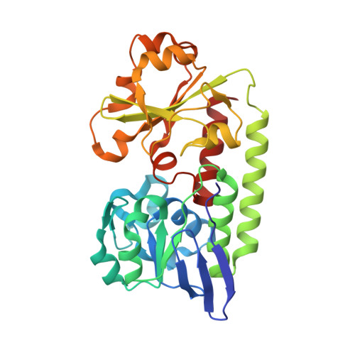
    Bacteria use siderophores to mediate the transport of essential Fe(III) into the cell. In Campylobacter jejuni the periplasmic binding protein CeuE, an integral part of the Fe(III) transport system, has adapted to bind tetradentate siderophores using a His and a Tyr side chain to complete the Fe(III) coordination. A series of tetradentate siderophore mimics was synthesized in which the length of the linker between the two iron-binding catecholamide units was increased from four carbon atoms (4-LICAM 4- ) to five, six and eight (5-, 6-, 8-LICAM 4- , respectively). Co-crystal structures with CeuE showed that the inter-planar angles between the iron-binding catecholamide units in the 5-, 6- and 8-LICAM 4- structures are very similar (111°, 110° and 110°) and allow for an optimum fit into the binding pocket of CeuE, the inter-planar angle in the structure of 4-LICAM 4- is significantly smaller (97°) due to restrictions imposed by the shorter linker. Accordingly, the protein-binding affinity was found to be slightly higher for 5- compared to 4-LICAM 4- but decreases for 6- and 8-LICAM 4- . The optimum linker length of five matches that present in natural siderophores such as enterobactin and azotochelin. Site-directed mutagenesis was used to investigate the relative importance of the Fe(III)-coordinating residues H227 and Y288.


  • Organizational Affiliation
    • Structural Biology Laboratory, Department of Chemistry, University of York, Heslington, York YO10 5DD, UK.

Macromolecule Content 

  • Total Structure Weight: 32.5 kDa 
  • Atom Count: 2,729 
  • Modeled Residue Count: 290 
  • Deposited Residue Count: 290 
  • Unique protein chains: 1

Macromolecules

Find similar proteins by:|  3D Structure
Entity ID: 1
MoleculeChains  Sequence LengthOrganismDetailsImage
ENTEROCHELIN UPTAKE PERIPLASMIC BINDING PROTEIN290Campylobacter jejuniMutation(s): 0 
UniProt
Find proteins for Q0P8Q4 (Campylobacter jejuni subsp. jejuni serotype O:2 (strain ATCC 700819 / NCTC 11168))
Explore Q0P8Q4 
Go to UniProtKB:  Q0P8Q4
Entity Groups
Sequence Clusters30% Identity50% Identity70% Identity90% Identity95% Identity100% Identity
UniProt GroupQ0P8Q4
Sequence Annotations
Expand
Reference Sequence

Small Molecules

Ligands 2 Unique
IDChains Name / Formula / InChI Key2D Diagram3D Interactions
8LC

Query on 8LC


Download:Ideal Coordinates CCD File
C [auth A]N,N'-OCTANE-1,8-DIYLBIS(2,3-DIHYDROXYBENZAMIDE)
C22 H28 N2 O6
DREKRZFIOMALFJ-UHFFFAOYSA-N
FE

Query on FE


Download:Ideal Coordinates CCD File
B [auth A]FE (III) ION
Fe
VTLYFUHAOXGGBS-UHFFFAOYSA-N

Experimental Data & Validation

Experimental Data

  • Method: X-RAY DIFFRACTION
  • Resolution: 1.32 Å
  • R-Value Free:  0.181 (Depositor), 0.188 (DCC) 
  • R-Value Work:  0.140 (Depositor), 0.152 (DCC) 
  • R-Value Observed: 0.142 (Depositor) 
Space Group: P 21 21 21
Unit Cell:
Length ( Å )Angle ( ˚ )
a = 42.98α = 90
b = 55.98β = 90
c = 140.08γ = 90
Software Package:
Software NamePurpose
REFMACrefinement
xia2data reduction
PHASERphasing

Structure Validation

View Full Validation Report


Ligand Structure Quality Assessment 


Entry History 

Deposition Data

Revision History  (Full details and data files)

  • Version 1.0: 2016-09-28
    Type: Initial release
  • Version 1.1: 2017-04-12
    Changes: Database references
  • Version 1.2: 2017-04-19
    Changes: Database references
  • Version 1.3: 2024-01-10
    Changes: Data collection, Database references, Derived calculations, Other, Refinement description