5A03 | pdb_00005a03

Crystal structure of aldose-aldose oxidoreductase from Caulobacter crescentus complexed with xylose


Experimental Data Snapshot

  • Method: X-RAY DIFFRACTION
  • Resolution: 1.85 Å
  • R-Value Free: 
    0.195 (Depositor), 0.204 (DCC) 
  • R-Value Work: 
    0.167 (Depositor), 0.178 (DCC) 
  • R-Value Observed: 
    0.168 (Depositor) 

Starting Model: experimental
View more details

wwPDB Validation 3D Report Full Report

Validation slider image for 5A03

Ligand Structure Quality Assessment 


This is version 2.1 of the entry. See complete history

Literature

Structure and Function of Caulobacter Crescentus Aldose-Aldose Oxidoreductase.

Taberman, H.Andberg, M.Koivula, A.Hakulinen, N.Penttila, M.Rouvinen, J.Parkkinen, T.

(2015) Biochem J 472: 297

  • DOI: https://doi.org/10.1042/BJ20150681
  • Primary Citation Related Structures: 
    5A02, 5A03, 5A04, 5A05, 5A06

  • PubMed Abstract: 

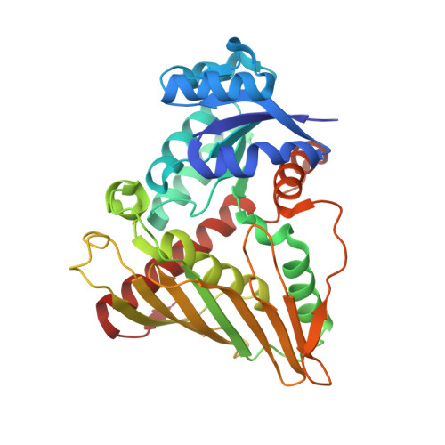
    Aldose-aldose oxidoreductase (Cc AAOR) is a recently characterized enzyme from the bacterial strain Caulobacter crescentus CB15 belonging to the glucose-fructose oxidoreductase/inositol dehydrogenase/rhizopine catabolism protein (Gfo/Idh/MocA) family. Cc AAOR catalyses the oxidation and reduction of a panel of aldose monosaccharides using a tightly bound NADP(H) cofactor that is regenerated in the catalytic cycle. Furthermore, Cc AAOR can also oxidize 1,4-linked oligosaccharides. In the present study, we present novel crystal structures of the dimeric Cc AAOR in complex with the cofactor and glycerol, D-xylose, D-glucose, maltotriose and D-sorbitol determined to resolutions of 2.0, 1.8, 1.7, 1.9 and 1.8 Å (1 Å=0.1 nm), respectively. These complex structures allowed for a detailed analysis of the ligand-binding interactions. The structures showed that the C1 carbon of a substrate, which is either reduced or oxidized, is close to the reactive C4 carbon of the nicotinamide ring of NADP(H). In addition, the O1 hydroxy group of the substrate, which is either protonated or deprotonated, is unexpectedly close to both Lys(104) and Tyr(189), which may both act as a proton donor or acceptor. This led us to hypothesize that this intriguing feature could be beneficial for Cc AAOR to catalyse the reduction of a linear form of a monosaccharide substrate and the oxidation of a pyranose form of the same substrate in a reaction cycle, during which the bound cofactor is regenerated.


  • Organizational Affiliation
    • Department of Chemistry, University of Eastern Finland, PO Box 111, FI-80101 Joensuu, Finland.

Macromolecule Content 

  • Total Structure Weight: 231.5 kDa 
  • Atom Count: 17,880 
  • Modeled Residue Count: 2,011 
  • Deposited Residue Count: 2,034 
  • Unique protein chains: 1

Macromolecules

Find similar proteins by:|  3D Structure
Entity ID: 1
MoleculeChains  Sequence LengthOrganismDetailsImage
ALDOSE-ALDOSE OXIDOREDUCTASE
A, B, C, D, E
A, B, C, D, E, F
339Caulobacter vibrioides CB15Mutation(s): 0 
EC: 1.1.99
UniProt
Find proteins for Q9A8X3 (Caulobacter vibrioides (strain ATCC 19089 / CIP 103742 / CB 15))
Explore Q9A8X3 
Go to UniProtKB:  Q9A8X3
Entity Groups
Sequence Clusters30% Identity50% Identity70% Identity90% Identity95% Identity100% Identity
UniProt GroupQ9A8X3
Sequence Annotations
Expand
Reference Sequence

Small Molecules

Ligands 5 Unique
IDChains Name / Formula / InChI Key2D Diagram3D Interactions
NDP

Query on NDP


Download:Ideal Coordinates CCD File
BA [auth E]
G [auth A]
JA [auth F]
N [auth B]
Q [auth C]
BA [auth E],
G [auth A],
JA [auth F],
N [auth B],
Q [auth C],
U [auth D]
NADPH DIHYDRO-NICOTINAMIDE-ADENINE-DINUCLEOTIDE PHOSPHATE
C21 H30 N7 O17 P3
ACFIXJIJDZMPPO-NNYOXOHSSA-N
XYS

Query on XYS


Download:Ideal Coordinates CCD File
FA [auth E],
P [auth B]
alpha-D-xylopyranose
C5 H10 O5
SRBFZHDQGSBBOR-LECHCGJUSA-N
XYP

Query on XYP


Download:Ideal Coordinates CCD File
CA [auth E]
DA [auth E]
EA [auth E]
H [auth A]
I [auth A]
CA [auth E],
DA [auth E],
EA [auth E],
H [auth A],
I [auth A],
J [auth A],
K [auth A],
KA [auth F],
O [auth B],
R [auth C],
S [auth C],
V [auth D],
W [auth D]
beta-D-xylopyranose
C5 H10 O5
SRBFZHDQGSBBOR-KKQCNMDGSA-N
SO4

Query on SO4


Download:Ideal Coordinates CCD File
AA [auth D]
GA [auth E]
HA [auth E]
IA [auth E]
L [auth A]
AA [auth D],
GA [auth E],
HA [auth E],
IA [auth E],
L [auth A],
LA [auth F],
M [auth A],
T [auth C],
Z [auth D]
SULFATE ION
O4 S
QAOWNCQODCNURD-UHFFFAOYSA-L
DIO

Query on DIO


Download:Ideal Coordinates CCD File
X [auth D],
Y [auth D]
1,4-DIETHYLENE DIOXIDE
C4 H8 O2
RYHBNJHYFVUHQT-UHFFFAOYSA-N

Experimental Data & Validation

Experimental Data

  • Method: X-RAY DIFFRACTION
  • Resolution: 1.85 Å
  • R-Value Free:  0.195 (Depositor), 0.204 (DCC) 
  • R-Value Work:  0.167 (Depositor), 0.178 (DCC) 
  • R-Value Observed: 0.168 (Depositor) 
Space Group: P 1 21 1
Unit Cell:
Length ( Å )Angle ( ˚ )
a = 101.059α = 90
b = 153.48β = 109.42
c = 107.327γ = 90
Software Package:
Software NamePurpose
PHENIXrefinement
XDSdata reduction
PHASERphasing

Structure Validation

View Full Validation Report


Ligand Structure Quality Assessment 


Entry History 

Deposition Data

Revision History  (Full details and data files)

  • Version 1.0: 2015-10-21
    Type: Initial release
  • Version 1.1: 2015-11-25
    Changes: Source and taxonomy
  • Version 1.2: 2015-12-09
    Changes: Database references
  • Version 2.0: 2020-07-29
    Type: Remediation
    Reason: Carbohydrate remediation
    Changes: Advisory, Atomic model, Data collection, Derived calculations, Other, Structure summary
  • Version 2.1: 2024-01-10
    Changes: Data collection, Database references, Refinement description, Structure summary