5ZYD | pdb_00005zyd

Crystal Structure of Glucose Isomerase Soaked with Glucose


Experimental Data Snapshot

  • Method: X-RAY DIFFRACTION
  • Resolution: 1.40 Å
  • R-Value Free: 
    0.165 (Depositor), 0.172 (DCC) 
  • R-Value Work: 
    0.151 (Depositor), 0.159 (DCC) 
  • R-Value Observed: 
    0.151 (Depositor) 

Starting Model: experimental
View more details

wwPDB Validation 3D Report Full Report

Validation slider image for 5ZYD

This is version 1.1 of the entry. See complete history

Literature

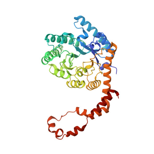
Structural analysis of substrate recognition by glucose isomerase in Mn2+binding mode at M2 site in S. rubiginosus

Bae, J.E.Hwang, K.Y.Nam, K.H.

(2018) Biochem Biophys Res Commun 503: 770-775

  • DOI: https://doi.org/10.1016/j.bbrc.2018.06.074
  • Primary Citation Related Structures: 
    5ZYC, 5ZYD, 5ZYE

  • PubMed Abstract: 

    Glucose isomerase (GI) catalyzes the reversible enzymatic isomerization of d-glucose and d-xylose to d-fructose and d-xylulose, respectively. This is one of the most important enzymes in the production of high-fructose corn syrup (HFCS) and biofuel. We recently determined the crystal structure of GI from S. rubiginosus (SruGI) complexed with a xylitol inhibitor in one metal binding mode. Although we assessed inhibitor binding at the M1 site, the metal binding at the M2 site and the substrate recognition mechanism for SruGI remains the unclear. Here, we report the crystal structure of the two metal binding modes of SruGI and its complex with glucose. This study provides a snapshot of metal binding at the SruGI M2 site in the presence of Mn 2+ , but not in the presence of Mg 2+ . Metal binding at the M2 site elicits a configuration change at the M1 site. Glucose molecule can only bind to the M1 site in presence of Mn 2+ at the M2 site. Glucose and Mn 2+ at the M2 site were bridged by water molecules using a hydrogen bonding network. The metal binding geometry of the M2 site indicates a distorted octahedral coordination with an angle of 55-110°, whereas the M1 site has a relatively stable octahedral coordination with an angle of 85-95°. We suggest a two-step sequential process for SruGI substrate recognition, in Mn 2+ binding mode, at the M2 site. Our results provide a better understanding of the molecular role of the M2 site in GI substrate recognition.


  • Organizational Affiliation
    • School of Life Sciences, KNU Creative BioResearch Group, Kyungpook National University, Daegu, Republic of Korea; KNU Institute for Microorganisms, Kyungpook National University, Daegu, Republic of Korea.

Macromolecule Content 

  • Total Structure Weight: 43.37 kDa 
  • Atom Count: 3,684 
  • Modeled Residue Count: 384 
  • Deposited Residue Count: 388 
  • Unique protein chains: 1

Macromolecules

Find similar proteins by:|  3D Structure
Entity ID: 1
MoleculeChains  Sequence LengthOrganismDetailsImage
Xylose isomerase388Streptomyces pseudogriseolusMutation(s): 0 
Gene Names: xylA
EC: 5.3.1.5
UniProt
Find proteins for P24300 (Streptomyces pseudogriseolus)
Explore P24300 
Go to UniProtKB:  P24300
Entity Groups
Sequence Clusters30% Identity50% Identity70% Identity90% Identity95% Identity100% Identity
UniProt GroupP24300
Sequence Annotations
Expand
Reference Sequence

Experimental Data & Validation

Experimental Data

  • Method: X-RAY DIFFRACTION
  • Resolution: 1.40 Å
  • R-Value Free:  0.165 (Depositor), 0.172 (DCC) 
  • R-Value Work:  0.151 (Depositor), 0.159 (DCC) 
  • R-Value Observed: 0.151 (Depositor) 
Space Group: I 2 2 2
Unit Cell:
Length ( Å )Angle ( ˚ )
a = 92.505α = 90
b = 97.82β = 90
c = 102.297γ = 90
Software Package:
Software NamePurpose
REFMACrefinement
PDB_EXTRACTdata extraction
HKL-2000data reduction
HKL-2000data scaling
MOLREPphasing

Structure Validation

View Full Validation Report


Entry History 

& Funding Information

Deposition Data

  • Released Date: 2018-11-28 
  • Deposition Author(s): Nam, K.H.

Funding OrganizationLocationGrant Number
National Research Foundation (Korea)Korea, Republic OfNRF-2017R1D1A1B03033087
National Research Foundation (Korea)Korea, Republic OfNRF-2017M3A9F6029736

Revision History  (Full details and data files)

  • Version 1.0: 2018-11-28
    Type: Initial release
  • Version 1.1: 2023-11-22
    Changes: Data collection, Database references, Refinement description