5ZXV | pdb_00005zxv

Structural definition of a unique neutralization epitope on the receptor-binding domain of MERS-CoV spike glycoprotein


Experimental Data Snapshot

  • Method: X-RAY DIFFRACTION
  • Resolution: 4.48 Å
  • R-Value Free: 
    0.343 (Depositor), 0.335 (DCC) 
  • R-Value Work: 
    0.307 (Depositor), 0.319 (DCC) 
  • R-Value Observed: 
    0.310 (Depositor) 

Starting Model: experimental
View more details

wwPDB Validation 3D Report Full Report

Validation slider image for 5ZXV

This is version 1.2 of the entry. See complete history

Literature

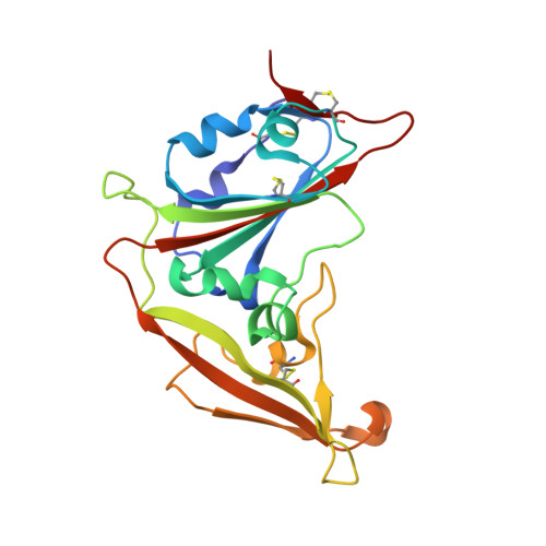
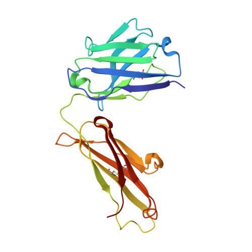
Structural Definition of a Unique Neutralization Epitope on the Receptor-Binding Domain of MERS-CoV Spike Glycoprotein

Zhang, S.Zhou, P.Wang, P.Li, Y.Jiang, L.Jia, W.Wang, H.Fan, A.Wang, D.Shi, X.Fang, X.Hammel, M.Wang, S.Wang, X.Zhang, L.

(2018) Cell Rep 24: 441-452

  • DOI: https://doi.org/10.1016/j.celrep.2018.06.041
  • Primary Citation Related Structures: 
    5YY5, 5ZXV

  • PubMed Abstract: 

    The major mechanism of antibody-mediated neutralization of the Middle East respiratory syndrome coronavirus (MERS-CoV) involves competition with the cellular receptor dipeptidyl peptidase 4 (DPP4) for binding to the receptor-binding domain (RBD) of the spike (S) glycoprotein. Here, we report a unique epitope and unusual neutralizing mechanism of the isolated human antibody MERS-4. Structurally, MERS-4 approached the RBD from the outside of the RBD-DPP4 binding interface. Such binding resulted in the folding of the β5-β6 loop toward a shallow groove on the RBD interface critical for accommodating DPP4. The key residues for binding are identified through site-directed mutagenesis. Structural modeling revealed that MERS-4 binds to RBD only in the "up" position in the S trimer. Furthermore, MERS-4 demonstrated synergy with several reported antibodies. These results indicate that MERS-4 neutralizes MERS-CoV by indirect rather than direct competition with DPP4. This mechanism provides a valuable addition for the combined use of antibodies against MERS-CoV infection.


  • Organizational Affiliation
    • The Ministry of Education Key Laboratory of Protein Science, Beijing Advanced Innovation Center for Structural Biology, Collaborative Innovation Center for Biotherapy, School of Life Sciences, Tsinghua University, Beijing 100084, China.

Macromolecule Content 

  • Total Structure Weight: 136.8 kDa 
  • Atom Count: 9,462 
  • Modeled Residue Count: 1,254 
  • Deposited Residue Count: 1,272 
  • Unique protein chains: 3

Macromolecules

Find similar proteins by:|  3D Structure
Entity ID: 1
MoleculeChains  Sequence LengthOrganismDetailsImage
MERS-CoV RBD
A, B
208Middle East respiratory syndrome-related coronavirusMutation(s): 0 
UniProt
Find proteins for K9N5Q8 (Middle East respiratory syndrome-related coronavirus (isolate United Kingdom/H123990006/2012))
Explore K9N5Q8 
Go to UniProtKB:  K9N5Q8
Entity Groups
Sequence Clusters30% Identity50% Identity70% Identity90% Identity95% Identity100% Identity
UniProt GroupK9N5Q8
Sequence Annotations
Expand
Reference Sequence
Find similar proteins by:|  3D Structure
Entity ID: 2
MoleculeChains  Sequence LengthOrganismDetailsImage
heavy chainC [auth H],
E [auth C]
213Homo sapiensMutation(s): 0 
Find similar proteins by:|  3D Structure
Entity ID: 3
MoleculeChains  Sequence LengthOrganismDetailsImage
light chainD [auth L],
F [auth D]
215Homo sapiensMutation(s): 0 

Experimental Data & Validation

Experimental Data

  • Method: X-RAY DIFFRACTION
  • Resolution: 4.48 Å
  • R-Value Free:  0.343 (Depositor), 0.335 (DCC) 
  • R-Value Work:  0.307 (Depositor), 0.319 (DCC) 
  • R-Value Observed: 0.310 (Depositor) 
Space Group: H 3
Unit Cell:
Length ( Å )Angle ( ˚ )
a = 138.647α = 90
b = 138.647β = 90
c = 228.736γ = 120
Software Package:
Software NamePurpose
PHENIXrefinement
HKL-2000data reduction
HKL-2000data scaling
PHASERphasing

Structure Validation

View Full Validation Report


Entry History 

& Funding Information

Deposition Data


Funding OrganizationLocationGrant Number
China--

Revision History  (Full details and data files)

  • Version 1.0: 2018-07-25
    Type: Initial release
  • Version 1.1: 2023-11-22
    Changes: Data collection, Database references, Refinement description
  • Version 1.2: 2024-10-16
    Changes: Structure summary