5ZX5 | pdb_00005zx5

3.3 angstrom structure of mouse TRPM7 with EDTA


Experimental Data Snapshot

  • Method: ELECTRON MICROSCOPY
  • Resolution: 3.28 Å
  • Aggregation State: PARTICLE 
  • Reconstruction Method: SINGLE PARTICLE 

wwPDB Validation 3D Report Full Report

Validation slider image for 5ZX5

This is version 1.3 of the entry. See complete history

Literature

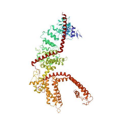
Structure of the mammalian TRPM7, a magnesium channel required during embryonic development.

Duan, J.Li, Z.Li, J.Hulse, R.E.Santa-Cruz, A.Valinsky, W.C.Abiria, S.A.Krapivinsky, G.Zhang, J.Clapham, D.E.

(2018) Proc Natl Acad Sci U S A 115: E8201-E8210

  • DOI: https://doi.org/10.1073/pnas.1810719115
  • Primary Citation Related Structures: 
    5ZX5, 6BWD, 6BWF

  • PubMed Abstract: 

    The transient receptor potential ion channel subfamily M, member 7 (TRPM7), is a ubiquitously expressed protein that is required for mouse embryonic development. TRPM7 contains both an ion channel and an α-kinase. The channel domain comprises a nonselective cation channel with notable permeability to Mg 2+ and Zn 2+ Here, we report the closed state structures of the mouse TRPM7 channel domain in three different ionic conditions to overall resolutions of 3.3, 3.7, and 4.1 Å. The structures reveal key residues for an ion binding site in the selectivity filter, with proposed partially hydrated Mg 2+ ions occupying the center of the conduction pore. In high [Mg 2+ ], a prominent external disulfide bond is found in the pore helix, which is essential for ion channel function. Our results provide a structural framework for understanding the TRPM1/3/6/7 subfamily and extend the knowledge base upon which to study the diversity and evolution of TRP channels.


  • Organizational Affiliation
    • Department of Cardiology, Boston Children's Hospital, Boston, MA 02115.

Macromolecule Content 

  • Total Structure Weight: 571.51 kDa 
  • Atom Count: 25,440 
  • Modeled Residue Count: 3,256 
  • Deposited Residue Count: 4,916 
  • Unique protein chains: 1

Macromolecules

Find similar proteins by:|  3D Structure
Entity ID: 1
MoleculeChains  Sequence LengthOrganismDetailsImage
Transient receptor potential cation channel subfamily M member 7
A, B, C, D
1,229Mus musculusMutation(s): 0 
Gene Names: Trpm7ChakLtrpc7
EC: 2.7.11.1
Membrane Entity: Yes 
UniProt & NIH Common Fund Data Resources
Find proteins for Q923J1 (Mus musculus)
Explore Q923J1 
Go to UniProtKB:  Q923J1
IMPC:  MGI:1929996
Entity Groups
Sequence Clusters30% Identity50% Identity70% Identity90% Identity95% Identity100% Identity
UniProt GroupQ923J1
Sequence Annotations
Expand
Reference Sequence

Small Molecules

Ligands 1 Unique
IDChains Name / Formula / InChI Key2D Diagram3D Interactions
Y01

Query on Y01


Download:Ideal Coordinates CCD File
E [auth A]
F [auth A]
G [auth A]
H [auth A]
I [auth A]
E [auth A],
F [auth A],
G [auth A],
H [auth A],
I [auth A],
J [auth B],
K [auth B],
L [auth C],
M [auth C],
N [auth D],
O [auth D],
P [auth D]
CHOLESTEROL HEMISUCCINATE
C31 H50 O4
WLNARFZDISHUGS-MIXBDBMTSA-N

Experimental Data & Validation

Experimental Data

  • Method: ELECTRON MICROSCOPY
  • Resolution: 3.28 Å
  • Aggregation State: PARTICLE 
  • Reconstruction Method: SINGLE PARTICLE 
EM Software:
TaskSoftware PackageVersion
MODEL REFINEMENTPHENIX

Structure Validation

View Full Validation Report


Entry History 

Deposition Data

Revision History  (Full details and data files)

  • Version 1.0: 2018-10-17
    Type: Initial release
  • Version 1.1: 2019-11-06
    Changes: Data collection, Other
  • Version 1.2: 2024-03-27
    Changes: Data collection, Database references
  • Version 1.3: 2025-06-18
    Changes: Data collection, Structure summary