5ZWF | pdb_00005zwf

Covalent bond formation between histidine of Vitamin D receptor (VDR) and a full agonist having a enone with a beta methyl group via conjugate addition reaction


Experimental Data Snapshot

  • Method: X-RAY DIFFRACTION
  • Resolution: 2.10 Å
  • R-Value Free: 
    0.250 (Depositor), 0.250 (DCC) 
  • R-Value Work: 
    0.234 (Depositor), 0.240 (DCC) 
  • R-Value Observed: 
    0.235 (Depositor) 

wwPDB Validation   3D Report Full Report


Ligand Structure Quality Assessment 

Created with Raphaël 2.3.0Worse 01 BetterLigand structure goodness of fit to experimental dataBest fitted 9KRClick on this verticalbar to view details

This is version 1.2 of the entry. See complete history


Literature

Identification of the Histidine Residue in Vitamin D Receptor That Covalently Binds to Electrophilic Ligands

Yoshizawa, M.Itoh, T.Hori, T.Kato, A.Anami, Y.Yoshimoto, N.Yamamoto, K.

(2018) J Med Chem 61: 6339-6349

  • DOI: https://doi.org/10.1021/acs.jmedchem.8b00774
  • Primary Citation of Related Structures:  
    5ZWE, 5ZWF, 5ZWH, 5ZWI

  • PubMed Abstract: 

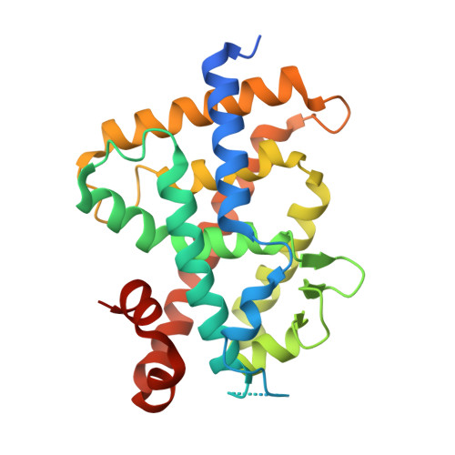
    We designed and synthesized vitamin D analogues with an electrophile as covalent modifiers for the vitamin D receptor (VDR). Novel vitamin D analogues 1-4 have an electrophilic enone group at the side chain for conjugate addition to His301 or His393 in the VDR. All compounds showed specific VDR-binding potency and agonistic activity. Covalent bond formations of 1-4 with the ligand-binding domain (LBD) of VDR were evaluated by electrospray ionization mass spectrometry. All compounds were shown to covalently bind to the VDR-LBD, and the abundance of VDR-LBD corresponding conjugate adducts of 1-4 increased with incubation time. Enone compounds 1 and 2 showed higher reactivity than the ene-ynone 3 and dienone 4 compounds. Furthermore, we successfully obtained cocrystals of VDR-LBD with analogues 1-4. X-ray crystallographic analysis showed a covalent bond with His301 in VDR-LBD. We successfully synthesized vitamin D analogues that form a covalent bond with VDR-LBD.


  • Organizational Affiliation

    Laboratory of Drug Design and Medicinal Chemistry , Showa Pharmaceutical University , 3-3165 Higashi-Tamagawagakuen , Machida, Tokyo 194-8543 , Japan.


Macromolecules
Find similar proteins by:  (by identity cutoff)  |  3D Structure
Entity ID: 1
MoleculeChains Sequence LengthOrganismDetailsImage
Vitamin D3 receptor271Rattus norvegicusMutation(s): 0 
Gene Names: VdrNr1i1
UniProt
Find proteins for P13053 (Rattus norvegicus)
Explore P13053 
Go to UniProtKB:  P13053
Entity Groups  
Sequence Clusters30% Identity50% Identity70% Identity90% Identity95% Identity100% Identity
UniProt GroupP13053
Sequence Annotations
Expand
  • Reference Sequence

Find similar proteins by:  Sequence   |   3D Structure  

Entity ID: 2
MoleculeChains Sequence LengthOrganismDetailsImage
13-meric peptide from DRIP205 NR2 BOX peptideB [auth C]13Homo sapiensMutation(s): 0 
UniProt & NIH Common Fund Data Resources
Find proteins for Q15648 (Homo sapiens)
Explore Q15648 
Go to UniProtKB:  Q15648
PHAROS:  Q15648
GTEx:  ENSG00000125686 
Entity Groups  
Sequence Clusters30% Identity50% Identity70% Identity90% Identity95% Identity100% Identity
UniProt GroupQ15648
Sequence Annotations
Expand
  • Reference Sequence
Small Molecules
Ligands 1 Unique
IDChains Name / Formula / InChI Key2D Diagram3D Interactions
9KR
Query on 9KR

Download Ideal Coordinates CCD File 
C [auth A](E,7R)-7-[(1R,3aS,4E,7aR)-7a-methyl-4-[2-[(3R,5R)-4-methylidene-3,5-bis(oxidanyl)cyclohexylidene]ethylidene]-2,3,3a,5,6,7-hexahydro-1H-inden-1-yl]oct-2-en-4-one
C27 H40 O3
VGMKRRGYTONKLW-FOWSJJJVSA-N
Experimental Data & Validation

Experimental Data

  • Method: X-RAY DIFFRACTION
  • Resolution: 2.10 Å
  • R-Value Free:  0.250 (Depositor), 0.250 (DCC) 
  • R-Value Work:  0.234 (Depositor), 0.240 (DCC) 
  • R-Value Observed: 0.235 (Depositor) 
Space Group: C 1 2 1
Unit Cell:
Length ( Å )Angle ( ˚ )
a = 153.56α = 90
b = 41.73β = 96.22
c = 42.02γ = 90
Software Package:
Software NamePurpose
REFMACrefinement
MOSFLMdata reduction
SCALAdata scaling
REFMACphasing

Structure Validation

View Full Validation Report


Ligand Structure Quality Assessment 

Created with Raphaël 2.3.0Worse 01 BetterLigand structure goodness of fit to experimental dataBest fitted 9KRClick on this verticalbar to view details

Entry History & Funding Information

Deposition Data


Funding OrganizationLocationGrant Number
Japan--

Revision History  (Full details and data files)

  • Version 1.0: 2018-07-18
    Type: Initial release
  • Version 1.1: 2018-08-08
    Changes: Data collection, Database references
  • Version 1.2: 2024-11-20
    Changes: Data collection, Database references, Structure summary