5ZSM | pdb_00005zsm

Crystal structure of monkey TLR7 in complex with GGUCCC


Experimental Data Snapshot

  • Method: X-RAY DIFFRACTION
  • Resolution: 2.50 Å
  • R-Value Free: 
    0.273 (Depositor), 0.272 (DCC) 
  • R-Value Work: 
    0.228 (Depositor), 0.233 (DCC) 
  • R-Value Observed: 
    0.231 (Depositor) 

wwPDB Validation 3D Report Full Report

Validation slider image for 5ZSM

Ligand Structure Quality Assessment 


This is version 2.2 of the entry. See complete history

Literature

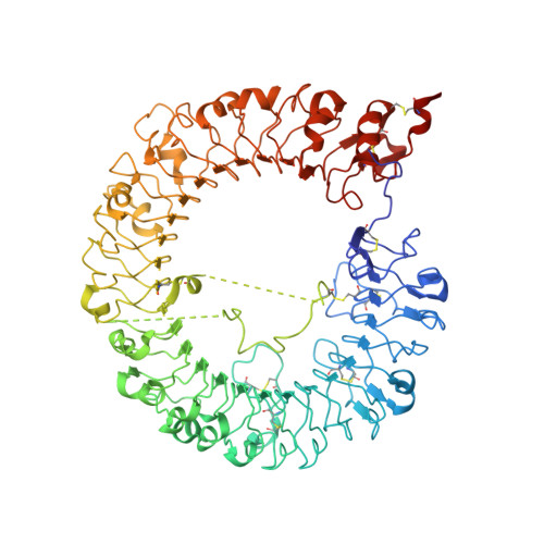
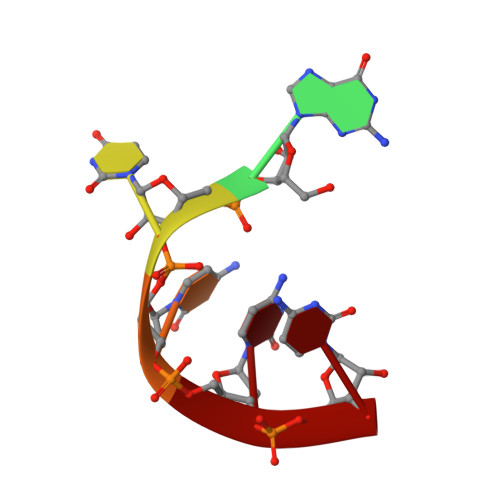
Structural Analyses of Toll-like Receptor 7 Reveal Detailed RNA Sequence Specificity and Recognition Mechanism of Agonistic Ligands.

Zhang, Z.Ohto, U.Shibata, T.Taoka, M.Yamauchi, Y.Sato, R.Shukla, N.M.David, S.A.Isobe, T.Miyake, K.Shimizu, T.

(2018) Cell Rep 25: 3371-3381.e5

  • DOI: https://doi.org/10.1016/j.celrep.2018.11.081
  • Primary Citation Related Structures: 
    5ZSA, 5ZSB, 5ZSC, 5ZSD, 5ZSE, 5ZSF, 5ZSG, 5ZSH, 5ZSI, 5ZSJ, 5ZSL, 5ZSM, 5ZSN, 6IF5

  • PubMed Abstract: 

    Toll-like receptor 7 (TLR7) is an innate immune receptor for single-stranded RNA (ssRNA) and has important roles in infectious diseases. We previously reported that TLR7 shows synergistic activation in response to two ligands, guanosine and ssRNA. However, the specific ssRNA sequence preference, detailed recognition mode of TLR7 and its ligand, and molecular determinants of TLR7 and TLR8 selectivity remain unknown. Here, we report on TLR7 from a large-scale crystallographic study combined with a multifaceted approach. We reveal that successive uridine-containing ssRNAs fully or moderately bind TLR7, whereas single uridine-containing ssRNAs have reduced affinities. We also reveal the detailed relationships between the chemical structures of ligands and their binding to TLR7. We demonstrate that an engineered TLR8 mutant alters its responsiveness to TLR7-specific ligands. Finally, we identify guanosine 2',3'-cyclic phosphate (2',3'-cGMP) as a possible endogenous ligand for TLR7 with greater affinity than guanosine. The abundant structural information will facilitate future development of treatments targeting TLR7.


  • Organizational Affiliation
    • Graduate School of Pharmaceutical Sciences, The University of Tokyo, 7-3-1 Hongo, Bunkyo-ku, Tokyo 113-0033, Japan.

Macromolecule Content 

  • Total Structure Weight: 198.51 kDa 
  • Atom Count: 13,150 
  • Modeled Residue Count: 1,560 
  • Deposited Residue Count: 1,658 
  • Unique protein chains: 1
  • Unique nucleic acid chains: 1

Macromolecules


Find similar proteins by:|  3D Structure
Entity ID: 1
MoleculeChains  Sequence LengthOrganismDetailsImage
Toll-like receptor 7A [auth B],
B [auth A]
823Macaca mulattaMutation(s): 4 
Gene Names: TLR7
UniProt
Find proteins for B3Y653 (Macaca mulatta)
Explore B3Y653 
Go to UniProtKB:  B3Y653
Entity Groups
Sequence Clusters30% Identity50% Identity70% Identity90% Identity95% Identity100% Identity
UniProt GroupB3Y653
Glycosylation
Glycosylation Sites: 9
Sequence Annotations
Expand
Reference Sequence
Find similar nucleic acids by:  Sequence
Entity ID: 2
MoleculeChains LengthOrganismImage
RNA (5'-R(*GP*UP*CP*CP*C)-3')C [auth D],
D [auth E]
6synthetic construct
Sequence Annotations
Expand
Reference Sequence

Oligosaccharides

Help  
Entity ID: 3
MoleculeChains Length2D Diagram GlycosylationD Interactions
2-acetamido-2-deoxy-beta-D-glucopyranose-(1-4)-2-acetamido-2-deoxy-beta-D-glucopyranoseE [auth C],
F
2N-Glycosylation
Glycosylation Resources
GlyTouCan: G42666HT
GlyCosmos: G42666HT
GlyGen: G42666HT

Small Molecules

Ligands 3 Unique
IDChains Name / Formula / InChI Key2D Diagram3D Interactions
9K9

Query on 9K9


Download:Ideal Coordinates CCD File
DA [auth A],
R [auth B]
2-amino-9-[(2S,3aR,4R,6R,6aR)-2-hydroxy-6-(hydroxymethyl)-2-oxotetrahydro-2H-2lambda~5~-furo[3,4-d][1,3,2]dioxaphosphol-4-yl]-3,9-dihydro-6H-purin-6-one
C10 H12 N5 O7 P
UASRYODFRYWBRC-UUOKFMHZSA-N
NAG

Query on NAG


Download:Ideal Coordinates CCD File
G [auth B]
H [auth B]
I [auth B]
J [auth B]
K [auth B]
G [auth B],
H [auth B],
I [auth B],
J [auth B],
K [auth B],
L [auth B],
M [auth B],
N [auth B],
S [auth A],
T [auth A],
U [auth A],
V [auth A],
W [auth A]
2-acetamido-2-deoxy-beta-D-glucopyranose
C8 H15 N O6
OVRNDRQMDRJTHS-FMDGEEDCSA-N
SO4

Query on SO4


Download:Ideal Coordinates CCD File
AA [auth A]
BA [auth A]
CA [auth A]
O [auth B]
P [auth B]
AA [auth A],
BA [auth A],
CA [auth A],
O [auth B],
P [auth B],
Q [auth B],
X [auth A],
Y [auth A],
Z [auth A]
SULFATE ION
O4 S
QAOWNCQODCNURD-UHFFFAOYSA-L

Experimental Data & Validation

Experimental Data

  • Method: X-RAY DIFFRACTION
  • Resolution: 2.50 Å
  • R-Value Free:  0.273 (Depositor), 0.272 (DCC) 
  • R-Value Work:  0.228 (Depositor), 0.233 (DCC) 
  • R-Value Observed: 0.231 (Depositor) 
Space Group: P 21 21 21
Unit Cell:
Length ( Å )Angle ( ˚ )
a = 99.655α = 90
b = 139.173β = 90
c = 150.794γ = 90
Software Package:
Software NamePurpose
REFMACrefinement
XDSdata reduction
Aimlessdata scaling
MOLREPphasing

Structure Validation

View Full Validation Report


Ligand Structure Quality Assessment 


Entry History 

Deposition Data

Revision History  (Full details and data files)

  • Version 1.0: 2019-01-30
    Type: Initial release
  • Version 2.0: 2020-07-29
    Type: Remediation
    Reason: Carbohydrate remediation
    Changes: Atomic model, Data collection, Derived calculations, Structure summary
  • Version 2.1: 2024-03-13
    Changes: Data collection, Database references, Refinement description, Structure summary
  • Version 2.2: 2024-10-30
    Changes: Structure summary