5ZRU | pdb_00005zru

Crystal structure of Agl-KA catalytic domain


Experimental Data Snapshot

  • Method: X-RAY DIFFRACTION
  • Resolution: 1.83 Å
  • R-Value Free: 
    0.171 (Depositor), 0.171 (DCC) 
  • R-Value Work: 
    0.141 (Depositor), 0.141 (DCC) 
  • R-Value Observed: 
    0.143 (Depositor) 

wwPDB Validation 3D Report Full Report

Validation slider image for 5ZRU

This is version 1.2 of the entry. See complete history

Literature

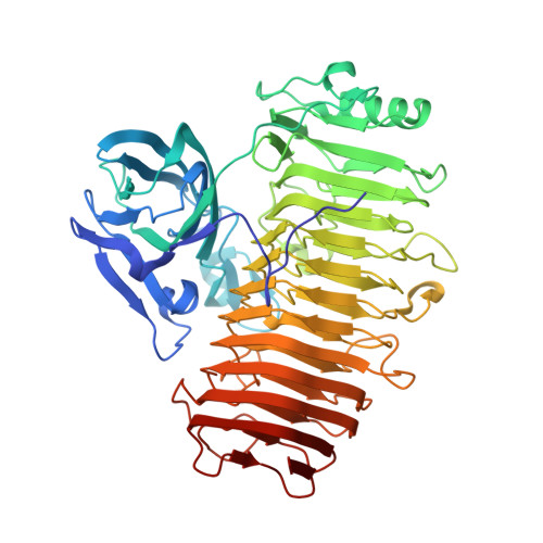
Crystal structure of the catalytic unit of GH 87-type alpha-1,3-glucanase Agl-KA from Bacillus circulans.

Yano, S.Suyotha, W.Oguro, N.Matsui, T.Shiga, S.Itoh, T.Hibi, T.Tanaka, Y.Wakayama, M.Makabe, K.

(2019) Sci Rep 9: 15295-15295

  • DOI: https://doi.org/10.1038/s41598-019-51822-5
  • Primary Citation Related Structures: 
    5ZRU

  • PubMed Abstract: 

    Glycoside hydrolase (GH) 87-type α-1,3-glucanase hydrolyses the α-1,3-glucoside linkages of α-1,3-glucan, which is found in fungal cell walls and extracellular polysaccharides produced by oral Streptococci. In this study, we report on the molecular structure of the catalytic unit of GH 87-type α-1,3-glucanase, Agl-KA, from Bacillus circulans, as determined by x-ray crystallography at a resolution of 1.82 Å. The catalytic unit constitutes a complex structure of two tandemly connected domains-the N-terminal galactose-binding-like domain and the C-terminal right-handed β-helix domain. While the β-helix domain is widely found among polysaccharide-processing enzymes, complex formation with the galactose-binding-like domain was observed for the first time. Biochemical assays showed that Asp1067, Asp1090 and Asp1091 are important for catalysis, and these residues are indeed located at the putative substrate-binding cleft, which forms a closed end and explains the product specificity.


  • Organizational Affiliation
    • Department of Biochemical Engineering, Graduate School of Sciences and Engineering, Yamagata University, Jonan, Yonezawa, Yamagata, 992-8510, Japan.

Macromolecule Content 

  • Total Structure Weight: 126.34 kDa 
  • Atom Count: 9,988 
  • Modeled Residue Count: 1,152 
  • Deposited Residue Count: 1,156 
  • Unique protein chains: 1

Macromolecules

Find similar proteins by:|  3D Structure
Entity ID: 1
MoleculeChains  Sequence LengthOrganismDetailsImage
Alpha-1,3-glucanaseA,
B [auth C]
578Niallia circulansMutation(s): 0 
Gene Names: agl
UniProt
Find proteins for Q0WYG9 (Niallia circulans)
Explore Q0WYG9 
Go to UniProtKB:  Q0WYG9
Entity Groups
Sequence Clusters30% Identity50% Identity70% Identity90% Identity95% Identity100% Identity
UniProt GroupQ0WYG9
Sequence Annotations
Expand
Reference Sequence

Small Molecules

Ligands 4 Unique
IDChains Name / Formula / InChI Key2D Diagram3D Interactions
PE5

Query on PE5


Download:Ideal Coordinates CCD File
C [auth A],
N [auth C]
3,6,9,12,15,18,21,24-OCTAOXAHEXACOSAN-1-OL
C18 H38 O9
CUDPPTPIUWYGFI-UHFFFAOYSA-N
SO4

Query on SO4


Download:Ideal Coordinates CCD File
D [auth A],
E [auth A],
O [auth C],
P [auth C]
SULFATE ION
O4 S
QAOWNCQODCNURD-UHFFFAOYSA-L
ZN

Query on ZN


Download:Ideal Coordinates CCD File
F [auth A],
G [auth A],
Q [auth C],
R [auth C]
ZINC ION
Zn
PTFCDOFLOPIGGS-UHFFFAOYSA-N
CA

Query on CA


Download:Ideal Coordinates CCD File
H [auth A]
I [auth A]
J [auth A]
K [auth A]
L [auth A]
H [auth A],
I [auth A],
J [auth A],
K [auth A],
L [auth A],
M [auth A],
S [auth C],
T [auth C],
U [auth C],
V [auth C]
CALCIUM ION
Ca
BHPQYMZQTOCNFJ-UHFFFAOYSA-N

Experimental Data & Validation

Experimental Data

  • Method: X-RAY DIFFRACTION
  • Resolution: 1.83 Å
  • R-Value Free:  0.171 (Depositor), 0.171 (DCC) 
  • R-Value Work:  0.141 (Depositor), 0.141 (DCC) 
  • R-Value Observed: 0.143 (Depositor) 
Space Group: P 1 21 1
Unit Cell:
Length ( Å )Angle ( ˚ )
a = 57.247α = 90
b = 126.423β = 97.83
c = 80.54γ = 90
Software Package:
Software NamePurpose
PHENIXrefinement
HKL-2000data reduction
HKL-2000data scaling
PHENIXphasing

Structure Validation

View Full Validation Report


Entry History 

Deposition Data

Revision History  (Full details and data files)

  • Version 1.0: 2019-10-23
    Type: Initial release
  • Version 1.1: 2020-05-06
    Changes: Database references
  • Version 1.2: 2024-03-27
    Changes: Data collection, Database references, Derived calculations, Structure summary