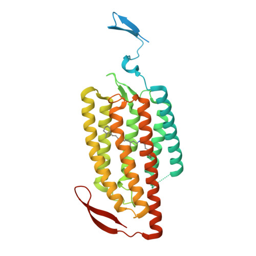
5ZIH

Crystal structure of the red light-activated channelrhodopsin Chrimson.


Experimental Data Snapshot

  • Method: X-RAY DIFFRACTION
  • Resolution: 2.60 Å
  • R-Value Free: 0.279 
  • R-Value Work: 0.228 
  • R-Value Observed: 0.233 

Starting Model: experimental
View more details

wwPDB Validation   3D Report Full Report


Ligand Structure Quality Assessment 


This is version 1.2 of the entry. See complete history


Literature

Crystal structure of the red light-activated channelrhodopsin Chrimson.

Oda, K.Vierock, J.Oishi, S.Rodriguez-Rozada, S.Taniguchi, R.Yamashita, K.Wiegert, J.S.Nishizawa, T.Hegemann, P.Nureki, O.

(2018) Nat Commun 9: 3949-3949

  • DOI: https://doi.org/10.1038/s41467-018-06421-9
  • Primary Citation of Related Structures:  
    5ZIH

  • PubMed Abstract: 

    Channelrhodopsins are light-activated ion channels that mediate cation permeation across cell membranes upon light absorption. Red-light-activated channelrhodopsins are of particular interest, because red light penetrates deeper into biological tissues and also enables dual-color experiments in combination with blue-light-activated optogenetic tools. Here we report the crystal structure of the most red-shifted channelrhodopsin from the algae Chlamydomonas noctigama, Chrimson, at 2.6 Å resolution. Chrimson resembles prokaryotic proton pumps in the retinal binding pocket, while sharing similarity with other channelrhodopsins in the ion-conducting pore. Concomitant mutation analysis identified the structural features that are responsible for Chrimson's red light sensitivity; namely, the protonation of the counterion for the retinal Schiff base, and the polar residue distribution and rigidity of the retinal binding pocket. Based on these mechanistic insights, we engineered ChrimsonSA, a mutant with a maximum activation wavelength red-shifted beyond 605 nm and accelerated closing kinetics.


  • Organizational Affiliation

    Department of Biological Sciences Graduate School of Science, The University of Tokyo, Tokyo, 113-0034, Japan.


Macromolecules
Find similar proteins by:  (by identity cutoff)  |  3D Structure
Entity ID: 1
MoleculeChains Sequence LengthOrganismDetailsImage
Sensory opsin A,Chrimson
A, B
347Chlamydomonas reinhardtiiChlamydomonas noctigama
This entity is chimeric
Mutation(s): 0 
Gene Names: CSOA
Membrane Entity: Yes 
UniProt
Find proteins for Q8L435 (Chlamydomonas reinhardtii)
Explore Q8L435 
Go to UniProtKB:  Q8L435
Entity Groups  
Sequence Clusters30% Identity50% Identity70% Identity90% Identity95% Identity100% Identity
UniProt GroupQ8L435
Sequence Annotations
Expand
  • Reference Sequence
Small Molecules
Ligands 1 Unique
IDChains Name / Formula / InChI Key2D Diagram3D Interactions
OLC
Query on OLC

Download Ideal Coordinates CCD File 
C [auth A]
D [auth A]
E [auth A]
F [auth A]
G [auth A]
C [auth A],
D [auth A],
E [auth A],
F [auth A],
G [auth A],
H [auth A],
I [auth A],
J [auth B],
K [auth B],
L [auth B],
M [auth B],
N [auth B],
O [auth B],
P [auth B],
Q [auth B],
R [auth B],
S [auth B],
T [auth B],
U [auth B]
(2R)-2,3-dihydroxypropyl (9Z)-octadec-9-enoate
C21 H40 O4
RZRNAYUHWVFMIP-GDCKJWNLSA-N
Modified Residues  1 Unique
IDChains TypeFormula2D DiagramParent
LYR
Query on LYR
A, B
L-PEPTIDE LINKINGC26 H42 N2 O2LYS
Experimental Data & Validation

Experimental Data

Unit Cell:
Length ( Å )Angle ( ˚ )
a = 60.99α = 90
b = 81.44β = 90
c = 170.14γ = 90
Software Package:
Software NamePurpose
XDSdata reduction
XSCALEdata scaling
PHENIXrefinement
PDB_EXTRACTdata extraction
PHASERphasing

Structure Validation

View Full Validation Report


Ligand Structure Quality Assessment 


Entry History & Funding Information

Deposition Data


Funding OrganizationLocationGrant Number
MEXTJapan16H06294
JSTJapanJPMJPR14L8

Revision History  (Full details and data files)

  • Version 1.0: 2018-11-21
    Type: Initial release
  • Version 1.1: 2018-11-28
    Changes: Data collection, Database references
  • Version 1.2: 2023-11-22
    Changes: Data collection, Database references, Refinement description