Primary Citation Related Structures: 5ZDP, 5ZDQ, 5ZDR
PubMed Abstract:
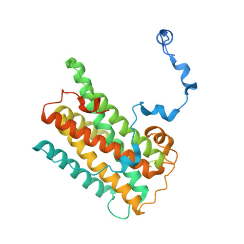
The alternative oxidase (AOX) is a monotopic diiron carboxylate protein which catalyzes the four-electron reduction of dioxygen to water by ubiquinol. Although we have recently determined the crystal structure of Trypanosoma brucei AOX (TAO) in the presence and absence of ascofuranone (AF) derivatives (which are potent mixed type inhibitors) the mechanism by which ubiquinol and dioxygen binds to TAO remain inconclusive. In this article, ferulenol was identified as the first competitive inhibitor of AOX which has been used to probe the binding of ubiquinol. Surface plasmon resonance reveals that AF is a quasi-irreversible inhibitor of TAO whilst ferulenol binding is completely reversible. The structure of the TAO-ferulenol complex, determined at 2.7 Å, provided insights into ubiquinol binding and has also identified a potential dioxygen molecule bound in a side-on conformation to the diiron center for the first time.
Organizational Affiliation:
Department of Applied Biology, Graduate School of Science and Technology, Kyoto Institute of Technology, Sakyo-ku, Matsugasaki, Kyoto 606-8585, Japan. Electronic address: tshiba@kit.ac.jp.
School of Tropical Medicine and Global Health, Nagasaki University, Sakamoto 1-12-4, Nagasaki 852-8523, Japan; Department of Host-Defense Biochemistry, Institute of Tropical Medicine (NEKKEN), Nagasaki University, Sakamoto 1-12-4, Nagasaki 852-8523, Japan; Department of Biomedical Chemistry, Graduate School of Medicine, The University of Tokyo, Bunkyo-ku, Hongo 7-3-1, Tokyo 113-0033, Japan. Electronic address: danielken@nagasaki-u.ac.jp.
Department of Applied Biology, Graduate School of Science and Technology, Kyoto Institute of Technology, Sakyo-ku, Matsugasaki, Kyoto 606-8585, Japan.
Department of Biomedical Chemistry, Graduate School of Medicine, The University of Tokyo, Bunkyo-ku, Hongo 7-3-1, Tokyo 113-0033, Japan.
School of Tropical Medicine and Global Health, Nagasaki University, Sakamoto 1-12-4, Nagasaki 852-8523, Japan; Department of Parasitology, Graduate School of Medicine, Osaka City University, Abeno-ku, Asahimachi 1-4-3, Osaka 545-8585, Japan.
Biochemistry and Biomedicine, School of Life Sciences, University of Sussex, Falmer, Brighton, BN1 9QG, UK.
Department of Applied Biology, Graduate School of Science and Technology, Kyoto Institute of Technology, Sakyo-ku, Matsugasaki, Kyoto 606-8585, Japan; Department of Biomedical Chemistry, Graduate School of Medicine, The University of Tokyo, Bunkyo-ku, Hongo 7-3-1, Tokyo 113-0033, Japan; Department of Biochemistry, Ahmadu Bello University, Zaria 2222, Nigeria.
Department of Molecular and Cellular Parasitology, Juntendo University School of Medicine, Bunkyo-ku, Hongo 2-1-1, Tokyo, 113-8421, Japan.
Systems and Structural Biology Center, RIKEN, Tsurumi, Suehiro 1-7-22, Yokohama, Kanagawa 230-0045, Japan.
Graduate School of Pharmaceutical Sciences, The University of Tokyo, Bunkyo-ku, Hongo 7-3-1, Tokyo 113-0033, Japan.
Department of Chemistry and Biotechnology, Graduate School of Engineering, Tottori University, Koyamacho-Minami 4, Tottori 680-8552, Japan.
School of Tropical Medicine and Global Health, Nagasaki University, Sakamoto 1-12-4, Nagasaki 852-8523, Japan; Department of Biomedical Chemistry, Graduate School of Medicine, The University of Tokyo, Bunkyo-ku, Hongo 7-3-1, Tokyo 113-0033, Japan; Department of Host-Defense Biochemistry, Institute of Tropical Medicine (NEKKEN), Nagasaki University, Sakamoto 1-12-4, Nagasaki 852-8523, Japan.