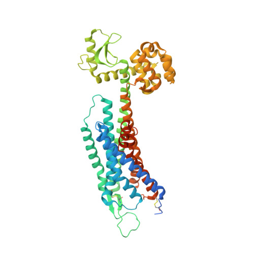
5ZBH

The Crystal Structure of Human Neuropeptide Y Y1 Receptor with BMS-193885


Experimental Data Snapshot

  • Method: X-RAY DIFFRACTION
  • Resolution: 3.00 Å
  • R-Value Free: 0.251 
  • R-Value Work: 0.222 
  • R-Value Observed: 0.224 

wwPDB Validation   3D Report Full Report


Ligand Structure Quality Assessment 


This is version 1.3 of the entry. See complete history


Literature

Structural basis of ligand binding modes at the neuropeptide Y Y1receptor

Yang, Z.Han, S.Keller, M.Kaiser, A.Bender, B.J.Bosse, M.Burkert, K.Kogler, L.M.Wifling, D.Bernhardt, G.Plank, N.Littmann, T.Schmidt, P.Yi, C.Li, B.Ye, S.Zhang, R.Xu, B.Larhammar, D.Stevens, R.C.Huster, D.Meiler, J.Zhao, Q.Beck-Sickinger, A.G.Buschauer, A.Wu, B.

(2018) Nature 556: 520-524

  • DOI: https://doi.org/10.1038/s41586-018-0046-x
  • Primary Citation of Related Structures:  
    5ZBH, 5ZBQ

  • PubMed Abstract: 

    Neuropeptide Y (NPY) receptors belong to the G-protein-coupled receptor superfamily and have important roles in food intake, anxiety and cancer biology 1,2 . The NPY-Y receptor system has emerged as one of the most complex networks with three peptide ligands (NPY, peptide YY and pancreatic polypeptide) binding to four receptors in most mammals, namely the Y 1 , Y 2 , Y 4 and Y 5 receptors, with different affinity and selectivity 3 . NPY is the most powerful stimulant of food intake and this effect is primarily mediated by the Y 1 receptor (Y 1 R) 4 . A number of peptides and small-molecule compounds have been characterized as Y 1 R antagonists and have shown clinical potential in the treatment of obesity 4 , tumour 1 and bone loss 5 . However, their clinical usage has been hampered by low potency and selectivity, poor brain penetration ability or lack of oral bioavailability 6 . Here we report crystal structures of the human Y 1 R bound to the two selective antagonists UR-MK299 and BMS-193885 at 2.7 and 3.0 Å resolution, respectively. The structures combined with mutagenesis studies reveal the binding modes of Y 1 R to several structurally diverse antagonists and the determinants of ligand selectivity. The Y 1 R structure and molecular docking of the endogenous agonist NPY, together with nuclear magnetic resonance, photo-crosslinking and functional studies, provide insights into the binding behaviour of the agonist and for the first time, to our knowledge, determine the interaction of its N terminus with the receptor. These insights into Y 1 R can enable structure-based drug discovery that targets NPY receptors.


  • Organizational Affiliation

    Chinese Academy of Sciences Key Laboratory of Receptor Research, Shanghai Institute of Materia Medica, Chinese Academy of Sciences, Shanghai, China.


Macromolecules
Find similar proteins by:  (by identity cutoff)  |  3D Structure
Entity ID: 1
MoleculeChains Sequence LengthOrganismDetailsImage
Neuropeptide Y receptor type 1,T4 Lysozyme,Neuropeptide Y receptor type 1527Homo sapiensEnterobacteria phage T4TMutation(s): 3 
Gene Names: NPY1RNPYRNPYY1eT4Tp126
EC: 3.2.1.17
Membrane Entity: Yes 
UniProt & NIH Common Fund Data Resources
Find proteins for P25929 (Homo sapiens)
Explore P25929 
Go to UniProtKB:  P25929
PHAROS:  P25929
GTEx:  ENSG00000164128 
Find proteins for P00720 (Enterobacteria phage T4)
Explore P00720 
Go to UniProtKB:  P00720
Entity Groups  
Sequence Clusters30% Identity50% Identity70% Identity90% Identity95% Identity100% Identity
UniProt GroupsP00720P25929
Sequence Annotations
Expand
  • Reference Sequence
Small Molecules
Ligands 1 Unique
IDChains Name / Formula / InChI Key2D Diagram3D Interactions
9AF
Query on 9AF

Download Ideal Coordinates CCD File 
B [auth A]dimethyl 4-{3-[({3-[4-(3-methoxyphenyl)piperidin-1-yl]propyl}carbamoyl)amino]phenyl}-2,6-dimethyl-1,4-dihydropyridine-3,5-dicarboxylate
C33 H42 N4 O6
WMYSXJSJXZFODY-UHFFFAOYSA-N
Binding Affinity Annotations 
IDSourceBinding Affinity
9AF BindingDB:  5ZBH Ki: 3.3 (nM) from 1 assay(s)
Experimental Data & Validation

Experimental Data

  • Method: X-RAY DIFFRACTION
  • Resolution: 3.00 Å
  • R-Value Free: 0.251 
  • R-Value Work: 0.222 
  • R-Value Observed: 0.224 
  • Space Group: C 2 2 21
Unit Cell:
Length ( Å )Angle ( ˚ )
a = 76.58α = 90
b = 126.24β = 90
c = 169.82γ = 90
Software Package:
Software NamePurpose
BUSTERrefinement
XDSdata reduction
XDSdata scaling
PHASERphasing

Structure Validation

View Full Validation Report


Ligand Structure Quality Assessment 


Entry History 

Deposition Data

Revision History  (Full details and data files)

  • Version 1.0: 2018-04-25
    Type: Initial release
  • Version 1.1: 2018-05-02
    Changes: Data collection, Database references
  • Version 1.2: 2018-05-09
    Changes: Data collection, Database references
  • Version 1.3: 2024-10-16
    Changes: Data collection, Database references, Structure summary