5Z65 | pdb_00005z65

Crystal structure of porcine aminopeptidase N ectodomain in functional form


Experimental Data Snapshot

  • Method: X-RAY DIFFRACTION
  • Resolution: 2.65 Å
  • R-Value Free: 
    0.251 (Depositor), 0.241 (DCC) 
  • R-Value Work: 
    0.196 (Depositor), 0.201 (DCC) 
  • R-Value Observed: 
    0.198 (Depositor) 

Starting Model: experimental
View more details

wwPDB Validation 3D Report Full Report

Validation slider image for 5Z65

Ligand Structure Quality Assessment 


This is version 2.2 of the entry. See complete history

Literature

Characterization of the interaction between recombinant porcine aminopeptidase N and spike glycoprotein of porcine epidemic diarrhea virus.

Sun, Y.G.Li, R.Jiang, L.Qiao, S.Zhi, Y.Chen, X.X.Xie, S.Wu, J.Li, X.Deng, R.Zhang, G.

(2018) Int J Biol Macromol 117: 704-712

  • DOI: https://doi.org/10.1016/j.ijbiomac.2018.05.167
  • Primary Citation Related Structures: 
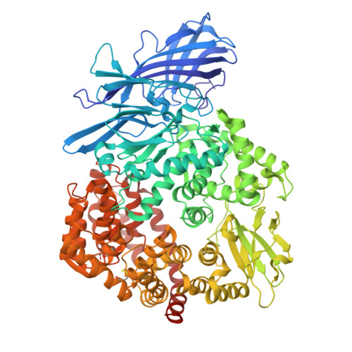
    5Z65

  • PubMed Abstract: 

    Porcine epidemic diarrhea (PED) has caused huge economic losses to the global pork industry. Infection by its causative agent PED virus (PEDV), an Alpha-coronavirus, was previously proven to be mediated by its spike (S) glycoprotein and a cellular receptor porcine aminopeptidase N (pAPN). Interestingly, some recent studies have indicated that pAPN is not a functional receptor for PEDV. To date, there is a lack of a direct evidence for the interaction between pAPN and PEDV S protein in vitro. Here, we prepared pAPN ectodomain and the truncated variants of PEDV S protein in Drosophila S2 cells. These recombinant proteins were homogeneous after purification by metal-affinity and size-exclusion chromatography. We then assayed the purified target proteins through immunogenicity tests, PEDV binding interference assays, circular dichroism (CD) measurements, pAPN activity assay and structural determination, demonstrating that they were biologically functional. Finally, we characterized their interactions by gel filtration chromatography, native-polyacrylamide gel electrophoresis (PAGE) and surface plasmon resonance (SPR) analyses. The results showed that their affinities were too low to form complexes, which suggest that pAPN may be controversial as the genuine receptor for PEDV. Therefore, further research needs to be carried out to elucidate the interaction between PEDV and its genuine receptor.


  • Organizational Affiliation
    • College of Veterinary Medicine, Jilin University, Changchun 130062, Jilin, China; Key Laboratory of Animal Immunology of the Ministry of Agriculture, Henan Provincial Key Laboratory of Animal Immunology, Henan Academy of Agricultural Sciences, Zhengzhou 450002, Henan, China.

Macromolecule Content 

  • Total Structure Weight: 108.21 kDa 
  • Atom Count: 7,530 
  • Modeled Residue Count: 900 
  • Deposited Residue Count: 913 
  • Unique protein chains: 1

Macromolecules

Find similar proteins by:|  3D Structure
Entity ID: 1
MoleculeChains  Sequence LengthOrganismDetailsImage
Aminopeptidase913Sus scrofaMutation(s): 0 
Gene Names: ANPEPpAPN
EC: 3.4.11 (PDB Primary Data), 3.4.11.2 (UniProt)
UniProt
Find proteins for P15145 (Sus scrofa)
Explore P15145 
Go to UniProtKB:  P15145
Entity Groups
Sequence Clusters30% Identity50% Identity70% Identity90% Identity95% Identity100% Identity
UniProt GroupP15145
Glycosylation
Glycosylation Sites: 9
Sequence Annotations
Expand
Reference Sequence

Oligosaccharides

Help  
Entity ID: 2
MoleculeChains Length2D Diagram GlycosylationD Interactions
2-acetamido-2-deoxy-beta-D-glucopyranose-(1-4)-2-acetamido-2-deoxy-beta-D-glucopyranose-(1-4)-2-acetamido-2-deoxy-beta-D-glucopyranose
B, C
3N-Glycosylation
Glycosylation Resources
GlyTouCan: G47362BJ
GlyCosmos: G47362BJ
GlyGen: G47362BJ
Entity ID: 3
MoleculeChains Length2D Diagram GlycosylationD Interactions
2-acetamido-2-deoxy-beta-D-glucopyranose-(1-4)-2-acetamido-2-deoxy-beta-D-glucopyranose
D, E, F, G, H
D, E, F, G, H, I
2N-Glycosylation
Glycosylation Resources
GlyTouCan: G42666HT
GlyCosmos: G42666HT
GlyGen: G42666HT

Small Molecules

Biologically Interesting Molecules (External Reference) 

1 Unique

Experimental Data & Validation

Experimental Data

  • Method: X-RAY DIFFRACTION
  • Resolution: 2.65 Å
  • R-Value Free:  0.251 (Depositor), 0.241 (DCC) 
  • R-Value Work:  0.196 (Depositor), 0.201 (DCC) 
  • R-Value Observed: 0.198 (Depositor) 
Space Group: C 1 2 1
Unit Cell:
Length ( Å )Angle ( ˚ )
a = 260.327α = 90
b = 62.31β = 100.06
c = 80.569γ = 90
Software Package:
Software NamePurpose
REFMACrefinement
HKL-2000data reduction
HKL-2000data scaling
MOLREPphasing

Structure Validation

View Full Validation Report


Ligand Structure Quality Assessment 


Entry History 

Deposition Data

Revision History  (Full details and data files)

  • Version 1.0: 2019-01-02
    Type: Initial release
  • Version 2.0: 2020-07-29
    Type: Remediation
    Reason: Carbohydrate remediation
    Changes: Advisory, Atomic model, Data collection, Derived calculations, Structure summary
  • Version 2.1: 2023-11-22
    Changes: Data collection, Database references, Refinement description, Structure summary
  • Version 2.2: 2024-11-13
    Changes: Structure summary