5Z1X | pdb_00005z1x

Crystal Structure of Laccase from Cerrena sp. RSD1


Experimental Data Snapshot

  • Method: X-RAY DIFFRACTION
  • Resolution: 1.38 Å
  • R-Value Free: 
    0.149 (Depositor), 0.150 (DCC) 
  • R-Value Work: 
    0.126 (Depositor), 0.126 (DCC) 
  • R-Value Observed: 
    0.127 (Depositor) 

Starting Model: experimental
View more details

wwPDB Validation 3D Report Full Report

Validation slider image for 5Z1X

Ligand Structure Quality Assessment 


This is version 2.2 of the entry. See complete history

Literature

Kinetic analysis and structural studies of a high-efficiency laccase fromCerrenasp. RSD1.

Wu, M.H.Lee, C.C.Hsiao, A.S.Yu, S.M.Wang, A.H.J.Ho, T.D.

(2018) FEBS Open Bio 8: 1230-1246

  • DOI: https://doi.org/10.1002/2211-5463.12459
  • Primary Citation Related Structures: 
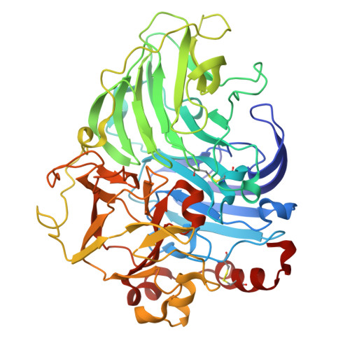
    5Z1X

  • PubMed Abstract: 

    A high-efficiency laccase, DLac, was isolated from Cerrena sp. RSD1. The kinetic studies indicate that DLac is a diffusion-limited enzyme. The crystal structure of DLac was determined to atomic resolution, and its overall structure shares high homology to monomeric laccases, but displays unique substrate-binding loops from those in other laccases. The substrate-binding residues with small side chain and the short substrate-binding loop IV broaden the substrate-binding cavity and may facilitate large substrate diffusion. Unlike highly glycosylated fungal laccases, the less-glycosylated DLac contains one highly conserved glycosylation site at N432 and an unique glycosylation site at N468. The N -glycans stabilize the substrate-binding loops and the protein structure, and the first N -acetylglucosamine is crucial for the catalytic efficiency. Additionally, a fivefold increase in protein yield is achieved via the submerged culture method for industrial applications. The atomic coordinates of the structure of DLac from Cerrena sp. RSD1 and structural factors have been deposited in the RCSB Protein Data Bank (PDB ID: 5Z1X).


  • Organizational Affiliation
    • Institute of Plant and Microbial Biology Academia Sinica Taipei Taiwan.

Macromolecule Content 

  • Total Structure Weight: 108.75 kDa 
  • Atom Count: 9,234 
  • Modeled Residue Count: 990 
  • Deposited Residue Count: 990 
  • Unique protein chains: 1

Macromolecules

Find similar proteins by:|  3D Structure
Entity ID: 1
MoleculeChains  Sequence LengthOrganismDetailsImage
Laccase
A, B
495CerrenaMutation(s): 0 
EC: 1.10.3.2
UniProt
Find proteins for A0A3F2YLU5 (Cerrena)
Explore A0A3F2YLU5 
Go to UniProtKB:  A0A3F2YLU5
Entity Groups
Sequence Clusters30% Identity50% Identity70% Identity90% Identity95% Identity100% Identity
UniProt GroupA0A3F2YLU5
Glycosylation
Glycosylation Sites: 2
Sequence Annotations
Expand
Reference Sequence

Oligosaccharides

Help  
Entity ID: 2
MoleculeChains Length2D Diagram GlycosylationD Interactions
2-acetamido-2-deoxy-beta-D-glucopyranose-(1-4)-2-acetamido-2-deoxy-beta-D-glucopyranose
C, D
2N-Glycosylation
Glycosylation Resources
GlyTouCan: G42666HT
GlyCosmos: G42666HT
GlyGen: G42666HT

Small Molecules

Ligands 5 Unique
IDChains Name / Formula / InChI Key2D Diagram3D Interactions
NAG

Query on NAG


Download:Ideal Coordinates CCD File
E [auth A],
P [auth B]
2-acetamido-2-deoxy-beta-D-glucopyranose
C8 H15 N O6
OVRNDRQMDRJTHS-FMDGEEDCSA-N
SO4

Query on SO4


Download:Ideal Coordinates CCD File
F [auth A]
G [auth A]
H [auth A]
Q [auth B]
R [auth B]
F [auth A],
G [auth A],
H [auth A],
Q [auth B],
R [auth B],
S [auth B]
SULFATE ION
O4 S
QAOWNCQODCNURD-UHFFFAOYSA-L
GOL

Query on GOL


Download:Ideal Coordinates CCD File
I [auth A],
J [auth A],
T [auth B],
U [auth B]
GLYCEROL
C3 H8 O3
PEDCQBHIVMGVHV-UHFFFAOYSA-N
CU

Query on CU


Download:Ideal Coordinates CCD File
K [auth A]
L [auth A]
M [auth A]
N [auth A]
V [auth B]
K [auth A],
L [auth A],
M [auth A],
N [auth A],
V [auth B],
W [auth B],
X [auth B],
Y [auth B]
COPPER (II) ION
Cu
JPVYNHNXODAKFH-UHFFFAOYSA-N
OXY

Query on OXY


Download:Ideal Coordinates CCD File
O [auth A],
Z [auth B]
OXYGEN MOLECULE
O2
MYMOFIZGZYHOMD-UHFFFAOYSA-N

Experimental Data & Validation

Experimental Data

  • Method: X-RAY DIFFRACTION
  • Resolution: 1.38 Å
  • R-Value Free:  0.149 (Depositor), 0.150 (DCC) 
  • R-Value Work:  0.126 (Depositor), 0.126 (DCC) 
  • R-Value Observed: 0.127 (Depositor) 
Space Group: P 21 21 21
Unit Cell:
Length ( Å )Angle ( ˚ )
a = 85.626α = 90
b = 88.795β = 90
c = 147.815γ = 90
Software Package:
Software NamePurpose
REFMACrefinement
HKL-2000data reduction
HKL-2000data scaling
MOLREPphasing

Structure Validation

View Full Validation Report


Ligand Structure Quality Assessment 


Entry History 

Deposition Data

Revision History  (Full details and data files)

  • Version 1.0: 2018-09-05
    Type: Initial release
  • Version 2.0: 2020-07-29
    Type: Remediation
    Reason: Carbohydrate remediation
    Changes: Atomic model, Data collection, Derived calculations, Structure summary
  • Version 2.1: 2023-11-22
    Changes: Data collection, Database references, Refinement description, Structure summary
  • Version 2.2: 2024-11-06
    Changes: Structure summary