5YWZ | pdb_00005ywz

AID-SUN tandem of SUN1


Experimental Data Snapshot

  • Method: X-RAY DIFFRACTION
  • Resolution: 2.20 Å
  • R-Value Free: 
    0.222 (Depositor), 0.227 (DCC) 
  • R-Value Work: 
    0.181 (Depositor), 0.187 (DCC) 
  • R-Value Observed: 
    0.183 (Depositor) 

Starting Model: experimental
View more details

wwPDB Validation 3D Report Full Report

Validation slider image for 5YWZ

This is version 1.2 of the entry. See complete history

Literature

Structural conservation of the autoinhibitory domain in SUN proteins

Xu, Y.Li, W.Ke, H.Feng, W.

(2018) Biochem Biophys Res Commun 496: 1337-1343

  • DOI: https://doi.org/10.1016/j.bbrc.2018.02.015
  • Primary Citation Related Structures: 
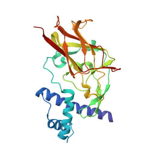
    5YWZ

  • PubMed Abstract: 

    LINC complexes span across the nuclear envelope and are assembled by SUN and KASH proteins. SUN1 and SUN2 are the two most abundant SUN proteins in mammals. In SUN2, the predicted coiled-coil domain preceding the SUN domain forms a three-helix bundle that constitutes an autoinhibitory domain (AID) to lock down the SUN domain. Here, we found that SUN1 also contains an AID preceding the SUN domain and solved the structure of the AID-SUN tandem of SUN1. SUN1 AID also adopts a three-helix bundle conformation that interacts with the SUN domain and keeps it in an autoinhibited state. Disruptions of the interaction interface in the AID-SUN tandem restored the SUN domain activity for binding to the KASH peptide. Structural comparison further demonstrated that the autoinhibited conformations of the AID-SUN tandems from SUN1 and SUN2 are similar and the intramolecular interdomain packing in SUN1 is slightly more compact than that in SUN2 due to minor variations of the residues in the interaction interface. Thus, AID is a conserved functional domain in SUN proteins and this work provides the structural evidence to support the conversation of the AID-mediated autoinhibition of SUN proteins.


  • Organizational Affiliation
    • National Laboratory of Biomacromolecules, CAS Center for Excellence in Biomacromolecules, Institute of Biophysics, Chinese Academy of Sciences, 15 Datun Road, Beijing 100101, China; University of Chinese Academy of Sciences, Beijing 100049, China.

Macromolecule Content 

  • Total Structure Weight: 27.08 kDa 
  • Atom Count: 2,090 
  • Modeled Residue Count: 242 
  • Deposited Residue Count: 242 
  • Unique protein chains: 1

Macromolecules

Find similar proteins by:|  3D Structure
Entity ID: 1
MoleculeChains  Sequence LengthOrganismDetailsImage
SUN domain-containing protein 1242Mus musculusMutation(s): 0 
Gene Names: Sun1Unc84a
UniProt & NIH Common Fund Data Resources
Find proteins for Q9D666 (Mus musculus)
Explore Q9D666 
Go to UniProtKB:  Q9D666
IMPC:  MGI:1924303
Entity Groups
Sequence Clusters30% Identity50% Identity70% Identity90% Identity95% Identity100% Identity
UniProt GroupQ9D666
Sequence Annotations
Expand
Reference Sequence

Experimental Data & Validation

Experimental Data

  • Method: X-RAY DIFFRACTION
  • Resolution: 2.20 Å
  • R-Value Free:  0.222 (Depositor), 0.227 (DCC) 
  • R-Value Work:  0.181 (Depositor), 0.187 (DCC) 
  • R-Value Observed: 0.183 (Depositor) 
Space Group: P 31 2 1
Unit Cell:
Length ( Å )Angle ( ˚ )
a = 73.636α = 90
b = 73.636β = 90
c = 97.456γ = 120
Software Package:
Software NamePurpose
HKL-2000data reduction
HKL-2000data scaling
PHASERphasing
PHENIXrefinement
PDB_EXTRACTdata extraction

Structure Validation

View Full Validation Report


Entry History 

& Funding Information

Deposition Data


Funding OrganizationLocationGrant Number
National Key R&D Program of ChinaChina2017YFA0503501

Revision History  (Full details and data files)

  • Version 1.0: 2018-02-21
    Type: Initial release
  • Version 1.1: 2018-02-28
    Changes: Database references
  • Version 1.2: 2023-11-22
    Changes: Data collection, Database references, Refinement description