5YRE | pdb_00005yre

Crystal structure of PPL3A


Experimental Data Snapshot

  • Method: X-RAY DIFFRACTION
  • Resolution: 1.40 Å
  • R-Value Free: 
    0.214 (Depositor), 0.217 (DCC) 
  • R-Value Work: 
    0.175 (Depositor), 0.177 (DCC) 
  • R-Value Observed: 
    0.177 (Depositor) 

Starting Model: experimental
View more details

wwPDB Validation 3D Report Full Report

Validation slider image for 5YRE

This is version 2.2 of the entry. See complete history

Literature

Structures of jacalin-related lectin PPL3 regulating pearl shell biomineralization

Nakae, S.Shionyu, M.Ogawa, T.Shirai, T.

(2018) Proteins 86: 644-653

  • DOI: https://doi.org/10.1002/prot.25491
  • Primary Citation Related Structures: 
    5YRE, 5YRF, 5YRG, 5YRH, 5YRI, 5YRJ, 5YRK, 5YRL, 5YRM

  • PubMed Abstract: 

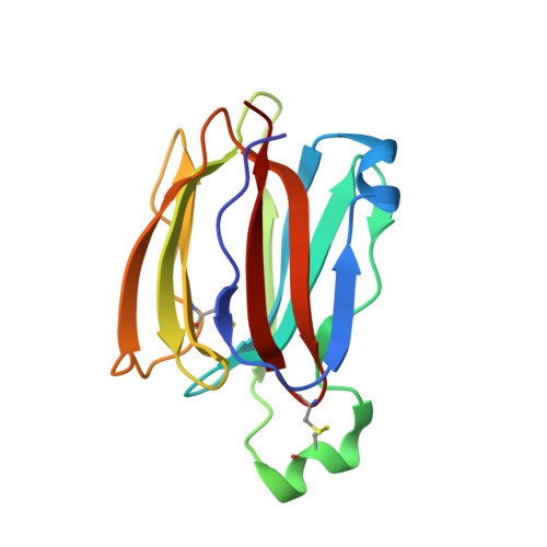
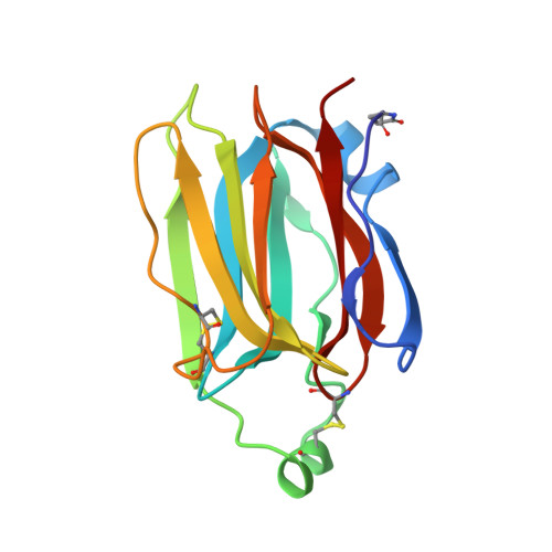
    The nacreous layer of pearl oysters is one of the major biominerals of commercial and industrial interest. Jacalin-related lectins, including PPL3 isoforms, are known to regulate biomineralization of the Pteria penguin pearl shell, although the molecular mechanisms are largely unknown. The PPL3 crystal structures were determined partly by utilizing microgravity environments for 3 isoforms, namely, PPL3A, PPL3B, and PPL3C. The structures revealed a tail-to-tail dimer structure established by forming a unique inter-subunit disulfide bond at C-termini. The N-terminal residues were found in pyroglutamate form, and this was partly explained by the post-translational modification of PPL3 isoforms implied from the discrepancy between amino acid and gene sequences. The complex structures with trehalose and isomaltose indicated that the novel specificity originated from the unique α-helix of PPL3 isoforms. Docking simulations of PPL3B to various calcite crystal faces suggested the edge of a β-sheet and the carbohydrate-binding site rich in charged residues were the interface to the biomineral, and implied that the isoforms differed in calcite interactions.


  • Organizational Affiliation
    • Faculty of Bioscience, Nagahama Institute of Bio-Science and Technology, 1266 Tamura-Cho, Nagahama, Shiga 526-0829, Japan.

Macromolecule Content 

  • Total Structure Weight: 32.33 kDa 
  • Atom Count: 2,714 
  • Modeled Residue Count: 284 
  • Deposited Residue Count: 284 
  • Unique protein chains: 2

Macromolecules

Find similar proteins by:|  3D Structure
Entity ID: 1
MoleculeChains  Sequence LengthOrganismDetailsImage
PPL3-A142Pteria penguinMutation(s): 0 
UniProt
Find proteins for B6F0T7 (Pteria penguin)
Explore B6F0T7 
Go to UniProtKB:  B6F0T7
Entity Groups
Sequence Clusters30% Identity50% Identity70% Identity90% Identity95% Identity100% Identity
UniProt GroupB6F0T7
Sequence Annotations
Expand
Reference Sequence
Find similar proteins by:|  3D Structure
Entity ID: 2
MoleculeChains  Sequence LengthOrganismDetailsImage
PPL3-A142Pteria penguinMutation(s): 0 
UniProt
Find proteins for B6F0T7 (Pteria penguin)
Explore B6F0T7 
Go to UniProtKB:  B6F0T7
Entity Groups
Sequence Clusters30% Identity50% Identity70% Identity90% Identity95% Identity100% Identity
UniProt GroupB6F0T7
Sequence Annotations
Expand
Reference Sequence

Small Molecules

Ligands 2 Unique
IDChains Name / Formula / InChI Key2D Diagram3D Interactions
SO4

Query on SO4


Download:Ideal Coordinates CCD File
C [auth A]
D [auth A]
E [auth A]
G [auth B]
H [auth B]
C [auth A],
D [auth A],
E [auth A],
G [auth B],
H [auth B],
I [auth B],
J [auth B]
SULFATE ION
O4 S
QAOWNCQODCNURD-UHFFFAOYSA-L
GOL

Query on GOL


Download:Ideal Coordinates CCD File
F [auth A],
K [auth B]
GLYCEROL
C3 H8 O3
PEDCQBHIVMGVHV-UHFFFAOYSA-N
Modified Residues  1 Unique
IDChains TypeFormula2D DiagramParent
PCA
Query on PCA
B
L-PEPTIDE LINKINGC5 H7 N O3GLN

Experimental Data & Validation

Experimental Data

  • Method: X-RAY DIFFRACTION
  • Resolution: 1.40 Å
  • R-Value Free:  0.214 (Depositor), 0.217 (DCC) 
  • R-Value Work:  0.175 (Depositor), 0.177 (DCC) 
  • R-Value Observed: 0.177 (Depositor) 
Space Group: P 21 21 21
Unit Cell:
Length ( Å )Angle ( ˚ )
a = 45.18α = 90
b = 50.21β = 90
c = 107.34γ = 90
Software Package:
Software NamePurpose
PHENIXrefinement
MOSFLMdata reduction
SCALAdata scaling
PHENIXphasing

Structure Validation

View Full Validation Report


Entry History 

& Funding Information

Deposition Data


Funding OrganizationLocationGrant Number
MEXTJapanJP17H01818
AMEDJapan17am0101069j0001
MEXTJapan#25107703
MEXTJapan#23107505

Revision History  (Full details and data files)

  • Version 1.0: 2018-08-29
    Type: Initial release
  • Version 2.0: 2020-01-01
    Changes: Database references, Derived calculations, Polymer sequence, Structure summary
  • Version 2.1: 2023-11-22
    Changes: Data collection, Database references, Refinement description
  • Version 2.2: 2024-11-06
    Changes: Structure summary