Targeting the potent Beclin 1-UVRAG coiled-coil interaction with designed peptides enhances autophagy and endolysosomal trafficking.
Wu, S., He, Y., Qiu, X., Yang, W., Liu, W., Li, X., Li, Y., Shen, H.M., Wang, R., Yue, Z., Zhao, Y.(2018) Proc Natl Acad Sci U S A 115: E5669-E5678
- PubMed: 29866835 Search on PubMedSearch on PubMed Central
- DOI: https://doi.org/10.1073/pnas.1721173115
- Primary Citation Related Structures:
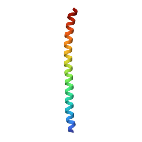
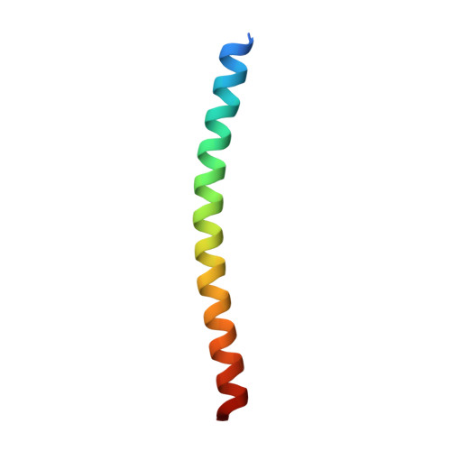
5YR0 - PubMed Abstract:
The Beclin 1-Vps34 complex, known as "mammalian class III PI3K," plays essential roles in membrane-mediated transport processes including autophagy and endosomal trafficking. Beclin 1 acts as a scaffolding molecule for the complex and readily transits from its metastable homodimeric state to interact with key modulators such as Atg14L or UVRAG and form functionally distinct Atg14L/UVRAG-containing Beclin 1-Vps34 subcomplexes. The Beclin 1-Atg14L/UVRAG interaction relies critically on their coiled-coil domains, but the molecular mechanism remains poorly understood. We determined the crystal structure of Beclin 1-UVRAG coiled-coil complex and identified a strengthened interface with both hydrophobic pairings and electrostatically complementary interactions. This structure explains why the Beclin 1-UVRAG interaction is more potent than the metastable Beclin 1 homodimer. Potent Beclin 1-UVRAG interaction is functionally significant because it renders UVRAG more competitive than Atg14L in Beclin 1 binding and is critical for promoting endolysosomal trafficking. UVRAG coiled-coil mutants with weakened Beclin 1 binding do not outcompete Atg14L and fail to promote endolysosomal degradation of the EGF receptor (EGFR). We designed all-hydrocarbon stapled peptides that specifically targeted the C-terminal part of the Beclin 1 coiled-coil domain to interfere with its homodimerization. One such peptide reduced Beclin 1 self-association, promoted Beclin 1-Atg14L/UVRAG interaction, increased autophagic flux, and enhanced EGFR degradation. Our results demonstrate that the targeting Beclin 1 coiled-coil domain with designed peptides to induce the redistribution of Beclin 1 among its self-associated form or Atg14L/UVRAG-containing complexes enhances both autophagy and endolysosomal trafficking.
- Department of Applied Biology and Chemical Technology, State Key Laboratory of Chirosciences, The Hong Kong Polytechnic University, Hung Hom, Kowloon, Hong Kong, People's Republic of China.
Organizational Affiliation: