5YOY | pdb_00005yoy

Crystal structure of the human tumor necrosis factor in complex with golimumab Fv


Experimental Data Snapshot

  • Method: X-RAY DIFFRACTION
  • Resolution: 2.73 Å
  • R-Value Free: 
    0.234 (Depositor), 0.236 (DCC) 
  • R-Value Work: 
    0.190 (Depositor), 0.197 (DCC) 
  • R-Value Observed: 
    0.192 (Depositor) 

Starting Models: experimental
View more details

wwPDB Validation 3D Report Full Report

Validation slider image for 5YOY

This is version 1.4 of the entry. See complete history

Literature

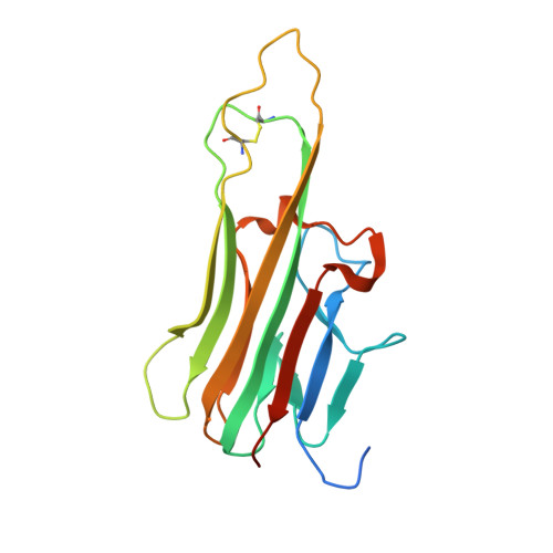
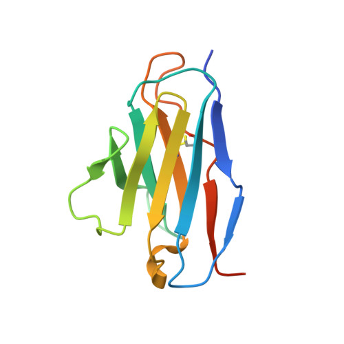
Structural basis for tumor necrosis factor blockade with the therapeutic antibody golimumab

Ono, M.Horita, S.Sato, Y.Nomura, Y.Iwata, S.Nomura, N.

(2018) Protein Sci 27: 1038-1046

  • DOI: https://doi.org/10.1002/pro.3407
  • Primary Citation Related Structures: 
    5YOY

  • PubMed Abstract: 

    Tumor necrosis factor α (TNFα) is a proinflammatory cytokine, and elevated levels of TNFα in serum are associated with various autoimmune diseases, including rheumatoid arthritis (RA), ankylosing spondylitis (AS), Crohn's disease (CD), psoriasis, and systemic lupus erythaematosus. TNFα performs its pleiotropic functions by binding to two structurally distinct transmembrane receptors, TNF receptor (TNFR) 1 and TNFR2. Antibody-based therapeutic strategies that block excessive TNFα signaling have been shown to be effective in suppressing such harmful inflammatory conditions. Golimumab (Simponi®) is an FDA-approved fully human monoclonal antibody targeting TNFα that has been widely used for the treatment of RA, AS, and CD. However, the structural basis underlying the inhibitory action of golimumab remains unclear. Here, we report the crystal structure of the Fv fragment of golimumab in complex with TNFα at a resolution of 2.73 Å. The resolved structure reveals that golimumab binds to a distinct epitope on TNFα that does not overlap with the binding residues of TNFR2. Golimumab exerts its inhibitory effect by preventing binding of TNFR1 and TNFR2 to TNFα by steric hindrance. Golimumab does not induce conformational changes in TNFα that could affect receptor binding. This mode of action is specific to golimumab among the four anti-TNFα therapeutic antibodies currently approved for clinical use.


  • Organizational Affiliation
    • Department of Cell Biology, Graduate School of Medicine, Kyoto University, Yoshida-Konoe-cho, Sakyo-ku, Kyoto, 606-8501, Japan.

Macromolecule Content 

  • Total Structure Weight: 279.32 kDa 
  • Atom Count: 18,116 
  • Modeled Residue Count: 2,324 
  • Deposited Residue Count: 2,526 
  • Unique protein chains: 3

Macromolecules

Find similar proteins by:|  3D Structure
Entity ID: 1
MoleculeChains  Sequence LengthOrganismDetailsImage
Tumor necrosis factor
A, B, C, J, K
A, B, C, J, K, L
173Homo sapiensMutation(s): 0 
Gene Names: TNFTNFATNFSF2
UniProt & NIH Common Fund Data Resources
Find proteins for P01375 (Homo sapiens)
Explore P01375 
Go to UniProtKB:  P01375
PHAROS:  P01375
GTEx:  ENSG00000232810 
Entity Groups
Sequence Clusters30% Identity50% Identity70% Identity90% Identity95% Identity100% Identity
UniProt GroupP01375
Sequence Annotations
Expand
Reference Sequence
Find similar proteins by:|  3D Structure
Entity ID: 2
MoleculeChains  Sequence LengthOrganismDetailsImage
Golimumab light chain variable region
D, E, F, M, N
D, E, F, M, N, O
119Homo sapiensMutation(s): 0 
Find similar proteins by:|  3D Structure
Entity ID: 3
MoleculeChains  Sequence LengthOrganismDetailsImage
Golimumab heavy chain variable region
G, H, I, P, Q
G, H, I, P, Q, R
129Homo sapiensMutation(s): 0 

Experimental Data & Validation

Experimental Data

  • Method: X-RAY DIFFRACTION
  • Resolution: 2.73 Å
  • R-Value Free:  0.234 (Depositor), 0.236 (DCC) 
  • R-Value Work:  0.190 (Depositor), 0.197 (DCC) 
  • R-Value Observed: 0.192 (Depositor) 
Space Group: P 1 21 1
Unit Cell:
Length ( Å )Angle ( ˚ )
a = 133.64α = 90
b = 85.3β = 101.61
c = 135.96γ = 90
Software Package:
Software NamePurpose
PHENIXrefinement
XSCALEdata scaling
PDB_EXTRACTdata extraction
XDSdata reduction
PHASERphasing

Structure Validation

View Full Validation Report


Entry History 

& Funding Information

Deposition Data


Funding OrganizationLocationGrant Number
Japan Science and TechnologyJapan--
Japan Society for the Promotion of ScienceJapan15K06968
Japan Society for the Promotion of ScienceJapan15J04343
Japan Agency of Medical Research and DevelopmentJapan--

Revision History  (Full details and data files)

  • Version 1.0: 2018-05-09
    Type: Initial release
  • Version 1.1: 2018-06-20
    Changes: Data collection, Database references
  • Version 1.2: 2018-11-14
    Changes: Data collection, Derived calculations
  • Version 1.3: 2023-11-22
    Changes: Data collection, Database references, Refinement description
  • Version 1.4: 2024-11-06
    Changes: Structure summary