5YKT | pdb_00005ykt

Crystal structure of a glutamate-1-semialdehyde-aminomutase (K286A) from Pseudomonas aeruginosa PAO1 in complex with PMP


Experimental Data Snapshot

  • Method: X-RAY DIFFRACTION
  • Resolution: 1.57 Å
  • R-Value Free: 
    0.180 (Depositor), 0.190 (DCC) 
  • R-Value Work: 
    0.154 (Depositor), 0.164 (DCC) 
  • R-Value Observed: 
    0.154 (Depositor) 

Starting Model: experimental
View more details

wwPDB Validation 3D Report Full Report

Validation slider image for 5YKT

Ligand Structure Quality Assessment 


This is version 1.1 of the entry. See complete history

Literature

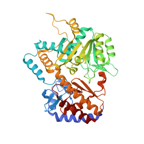
Crystal structure of a glutamate-1-semialdehyde-aminomutase from Pseudomonas aeruginosa PAO1.

Li, S.Lou, X.Xu, Y.Teng, X.Che, S.Liu, R.Bartlam, M.

(2018) Biochem Biophys Res Commun 500: 804-809

  • DOI: https://doi.org/10.1016/j.bbrc.2018.04.163
  • Primary Citation Related Structures: 
    5YKR, 5YKT

  • PubMed Abstract: 

    The C5 pathway in bacteria is responsible for the synthesis of 5-aminolevulinic acid, which forms the core skeleton of cofactors required for metabolism. One of the key actors in this pathway is a pyridoxamine-5'-phosphate (PMP)/pyridoxal-5'-phosphate (PLP) dependent enzyme called glutamate-1-semialdehyde aminomutase (GSAM). In this study, we crystallized the expression product of the uncharacterized pa4088 gene from the opportunistic pathogen Pseudomonas aeruginosa PAO1. The resulting high-resolution structure confirms it to be a member of the GSAM family. Continuous electron density indicates the presence of a PLP cofactor with a Schiff base linkage between the PLP cofactor and the ε-amino group of Lys286. A crystal structure of a K286A mutant in complex with PMP is also reported. As GSAM enzymes are not present in mammalian cells, this work provides a starting point for the investigation of GSAM as a target for drug development against P. aeruginosa infection.


  • Organizational Affiliation
    • College of Life Sciences, Nankai University, Tianjin, China.

Macromolecule Content 

  • Total Structure Weight: 102.74 kDa 
  • Atom Count: 7,960 
  • Modeled Residue Count: 906 
  • Deposited Residue Count: 922 
  • Unique protein chains: 1

Macromolecules

Find similar proteins by:|  3D Structure
Entity ID: 1
MoleculeChains  Sequence LengthOrganismDetailsImage
Probable aminotransferase
A, B
461Pseudomonas aeruginosa PAO1Mutation(s): 1 
Gene Names: PA4088
EC: 5.4.3.8
UniProt
Find proteins for Q9HWU0 (Pseudomonas aeruginosa (strain ATCC 15692 / DSM 22644 / CIP 104116 / JCM 14847 / LMG 12228 / 1C / PRS 101 / PAO1))
Explore Q9HWU0 
Go to UniProtKB:  Q9HWU0
Entity Groups
Sequence Clusters30% Identity50% Identity70% Identity90% Identity95% Identity100% Identity
UniProt GroupQ9HWU0
Sequence Annotations
Expand
Reference Sequence

Experimental Data & Validation

Experimental Data

  • Method: X-RAY DIFFRACTION
  • Resolution: 1.57 Å
  • R-Value Free:  0.180 (Depositor), 0.190 (DCC) 
  • R-Value Work:  0.154 (Depositor), 0.164 (DCC) 
  • R-Value Observed: 0.154 (Depositor) 
Space Group: P 21 21 21
Unit Cell:
Length ( Å )Angle ( ˚ )
a = 89.73α = 90
b = 97.104β = 90
c = 100.52γ = 90
Software Package:
Software NamePurpose
REFMACrefinement
HKL-2000data reduction
HKL-2000data scaling
PHASERphasing

Structure Validation

View Full Validation Report


Ligand Structure Quality Assessment 


Entry History 

& Funding Information

Deposition Data


Funding OrganizationLocationGrant Number
National Science Foundation (China)China31570128

Revision History  (Full details and data files)

  • Version 1.0: 2018-09-26
    Type: Initial release
  • Version 1.1: 2023-11-22
    Changes: Data collection, Database references, Refinement description