5YJG | pdb_00005yjg

Structural insights into periostin functions


Experimental Data Snapshot

  • Method: X-RAY DIFFRACTION
  • Resolution: 2.40 Å
  • R-Value Free: 
    0.247 (Depositor), 0.249 (DCC) 
  • R-Value Work: 
    0.211 (Depositor), 0.213 (DCC) 
  • R-Value Observed: 
    0.213 (Depositor) 

Starting Model: experimental
View more details

wwPDB Validation 3D Report Full Report

Validation slider image for 5YJG

This is version 1.4 of the entry. See complete history

Literature

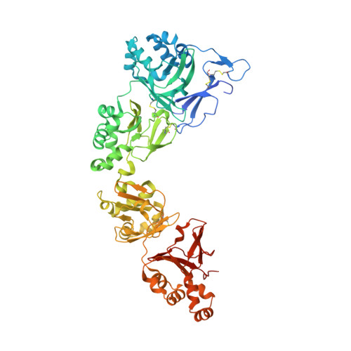
Structural characterizations of human periostin dimerization and cysteinylation.

Liu, J.Zhang, J.Xu, F.Lin, Z.Li, Z.Liu, H.

(2018) FEBS Lett 592: 1789-1803

  • DOI: https://doi.org/10.1002/1873-3468.13091
  • Primary Citation Related Structures: 
    5YJG, 5YJH

  • PubMed Abstract: 

    Human periostin plays a multifaceted role in remodeling the extracellular matrix milieu by interacting with other proteins and itself in both a heterophilic and homophilic manner. However, the structural mechanism for its extensive interactions has remained elusive. Here, we report the crystal structures of human periostin (EMI-Fas1 I- IV ) and its Cys60Ala mutant. In combination with multi-angle light-scattering analysis and biochemical assays, the crystal structures reveal that periostin mainly exists as a dimer in solution and its homophilic interaction is mainly mediated by the EMI domain. Furthermore, Cys60 undergoes cysteinylation as confirmed by mass spectroscopy, and this site hardly affects the homophilic interaction. Also, the structures yield insights into how periostin forms heterophilic interactions with other proteins under physiological or pathological conditions.


  • Organizational Affiliation
    • State Key Laboratory of Natural and Biomimetic Drugs, School of Pharmaceutical Sciences, Peking University Health Science Center, Haidian District, Beijing, China.

Macromolecule Content 

  • Total Structure Weight: 71.02 kDa 
  • Atom Count: 5,046 
  • Modeled Residue Count: 594 
  • Deposited Residue Count: 628 
  • Unique protein chains: 1

Macromolecules

Find similar proteins by:|  3D Structure
Entity ID: 1
MoleculeChains  Sequence LengthOrganismDetailsImage
Periostin628Homo sapiensMutation(s): 0 
Gene Names: POSTNOSF2
UniProt & NIH Common Fund Data Resources
Find proteins for Q15063 (Homo sapiens)
Explore Q15063 
Go to UniProtKB:  Q15063
PHAROS:  Q15063
GTEx:  ENSG00000133110 
Entity Groups
Sequence Clusters30% Identity50% Identity70% Identity90% Identity95% Identity100% Identity
UniProt GroupQ15063
Sequence Annotations
Expand
Reference Sequence

Small Molecules

Ligands 6 Unique
IDChains Name / Formula / InChI Key2D Diagram3D Interactions
CYS

Query on CYS


Download:Ideal Coordinates CCD File
C [auth A]CYSTEINE
C3 H7 N O2 S
XUJNEKJLAYXESH-REOHCLBHSA-N
ZN

Query on ZN


Download:Ideal Coordinates CCD File
HA [auth A]ZINC ION
Zn
PTFCDOFLOPIGGS-UHFFFAOYSA-N
CA

Query on CA


Download:Ideal Coordinates CCD File
T [auth A]
U [auth A]
V [auth A]
W [auth A]
X [auth A]
T [auth A],
U [auth A],
V [auth A],
W [auth A],
X [auth A],
Y [auth A]
CALCIUM ION
Ca
BHPQYMZQTOCNFJ-UHFFFAOYSA-N
CL

Query on CL


Download:Ideal Coordinates CCD File
D [auth A]
E [auth A]
F [auth A]
G [auth A]
H [auth A]
D [auth A],
E [auth A],
F [auth A],
G [auth A],
H [auth A],
I [auth A],
J [auth A],
K [auth A],
L [auth A],
M [auth A],
N [auth A],
O [auth A],
P [auth A],
Q [auth A],
R [auth A],
S [auth A]
CHLORIDE ION
Cl
VEXZGXHMUGYJMC-UHFFFAOYSA-M
MG

Query on MG


Download:Ideal Coordinates CCD File
AA [auth A]
BA [auth A]
CA [auth A]
DA [auth A]
EA [auth A]
AA [auth A],
BA [auth A],
CA [auth A],
DA [auth A],
EA [auth A],
FA [auth A],
GA [auth A],
Z [auth A]
MAGNESIUM ION
Mg
JLVVSXFLKOJNIY-UHFFFAOYSA-N
NA

Query on NA


Download:Ideal Coordinates CCD File
B [auth A]SODIUM ION
Na
FKNQFGJONOIPTF-UHFFFAOYSA-N

Experimental Data & Validation

Experimental Data

  • Method: X-RAY DIFFRACTION
  • Resolution: 2.40 Å
  • R-Value Free:  0.247 (Depositor), 0.249 (DCC) 
  • R-Value Work:  0.211 (Depositor), 0.213 (DCC) 
  • R-Value Observed: 0.213 (Depositor) 
Space Group: P 61 2 2
Unit Cell:
Length ( Å )Angle ( ˚ )
a = 105.605α = 90
b = 105.605β = 90
c = 331.225γ = 120
Software Package:
Software NamePurpose
PHENIXrefinement
HKL-3000data scaling

Structure Validation

View Full Validation Report


Entry History 

Deposition Data

Revision History  (Full details and data files)

  • Version 1.0: 2018-05-23
    Type: Initial release
  • Version 1.1: 2018-06-13
    Changes: Data collection, Database references
  • Version 1.2: 2018-06-20
    Changes: Data collection, Database references
  • Version 1.3: 2023-11-22
    Changes: Data collection, Database references, Derived calculations, Refinement description
  • Version 1.4: 2024-11-13
    Changes: Structure summary