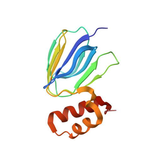
The NMR solution structure of Mycobacterium tuberculosis F-ATP synthase subunit epsilon provides new insight into energy coupling inside the rotary engine.
Joon, S., Ragunathan, P., Sundararaman, L., Nartey, W., Kundu, S., Manimekalai, M.S.S., Bogdanovic, N., Dick, T., Gruber, G.(2018) FEBS J 285: 1111-1128
- PubMed: 29360236 Search on PubMed
- DOI: https://doi.org/10.1111/febs.14392
- Primary Citation Related Structures:
5YIO - PubMed Abstract:
Mycobacterium tuberculosis (Mt) F 1 F 0 ATP synthase (α 3 :β 3 :γ:δ:ε:a:b:b':c 9 ) is essential for the viability of growing and nongrowing persister cells of the pathogen. Here, we present the first NMR solution structure of Mtε, revealing an N-terminal β-barrel domain (NTD) and a C-terminal domain (CTD) composed of a helix-loop-helix with helix 1 and -2 being shorter compared to their counterparts in other bacteria. The C-terminal amino acids are oriented toward the NTD, forming a domain-domain interface between the NTD and CTD. The Mtε structure provides a novel mechanistic model of coupling c-ring- and ε rotation via a patch of hydrophobic residues in the NTD and residues of the CTD to the bottom of the catalytic α 3 β 3 -headpiece. To test our model, genome site-directed mutagenesis was employed to introduce amino acid changes in these two parts of the epsilon subunit. Inverted vesicle assays show that these mutations caused an increase in ATP hydrolysis activity and a reduction in ATP synthesis. The structural and enzymatic data are discussed in light of the transition mechanism of a compact and extended state of Mtε, which provides the inhibitory effects of this coupling subunit inside the rotary engine. Finally, the employment of these data with molecular docking shed light into the second binding site of the drug Bedaquiline. Structural data are available in the PDB under the accession number 5YIO.
- Nanyang Technological University, School of Biological Sciences, Singapore, Singapore.
Organizational Affiliation: