5YEW | pdb_00005yew

Structural basis for GTP hydrolysis and conformational change of mitofusin 1 in mediating mitochondrial fusion


Experimental Data Snapshot

  • Method: X-RAY DIFFRACTION
  • Resolution: 3.20 Å
  • R-Value Free: 
    0.257 (Depositor), 0.256 (DCC) 
  • R-Value Work: 
    0.223 (Depositor), 0.222 (DCC) 
  • R-Value Observed: 
    0.224 (Depositor) 

wwPDB Validation 3D Report Full Report

Validation slider image for 5YEW

Ligand Structure Quality Assessment 


This is version 1.5 of the entry. See complete history

Literature

Structural basis for GTP hydrolysis and conformational change of MFN1 in mediating membrane fusion

Yan, L.Qi, Y.Huang, X.Yu, C.Lan, L.Guo, X.Rao, Z.Hu, J.Lou, Z.

(2018) Nat Struct Mol Biol 25: 233-243

  • DOI: https://doi.org/10.1038/s41594-018-0034-8
  • Primary Citation Related Structures: 
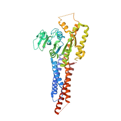
    5YEW

  • PubMed Abstract: 

    Fusion of the outer mitochondrial membrane is mediated by the dynamin-like GTPase mitofusin (MFN). Here, we determined the structure of the minimal GTPase domain (MGD) of human MFN1 in complex with GDP-BeF 3 - . The MGD folds into a canonical GTPase fold with an associating four-helix bundle, HB1, and forms a dimer. A potassium ion in the catalytic core engages GDP and BeF 3 - (GDP-BeF 3 - ). Enzymatic analysis has confirmed that efficient GTP hydrolysis by MFN1 requires potassium. Compared to previously reported MGD structures, the HB1 structure undergoes a major conformational change relative to the GTPase domains, as they move from pointing in opposite directions to point in the same direction, suggesting that a swing of the four-helix bundle can pull tethered membranes closer to achieve fusion. The proposed model is supported by results from in vitro biochemical assays and mitochondria morphology rescue assays in MFN1-deleted cells. These findings offer an explanation for how Charcot-Marie-Tooth neuropathy type 2 A (CMT2A)-causing mutations compromise MFN-mediated fusion.


  • Organizational Affiliation
    • School of Medicine, Tsinghua University, Beijing, China.

Macromolecule Content 

  • Total Structure Weight: 143.91 kDa 
  • Atom Count: 9,431 
  • Modeled Residue Count: 1,166 
  • Deposited Residue Count: 1,263 
  • Unique protein chains: 2

Macromolecules

Find similar proteins by:|  3D Structure
Entity ID: 1
MoleculeChains  Sequence LengthOrganismDetailsImage
Mitofusin-1,Mitofusin-1 fusion protein421Homo sapiensMutation(s): 2 
Gene Names: MFN1
EC: 3.6.5
UniProt & NIH Common Fund Data Resources
Find proteins for Q8IWA4 (Homo sapiens)
Explore Q8IWA4 
Go to UniProtKB:  Q8IWA4
PHAROS:  Q8IWA4
GTEx:  ENSG00000171109 
Entity Groups
Sequence Clusters30% Identity50% Identity70% Identity90% Identity95% Identity100% Identity
UniProt GroupQ8IWA4
Sequence Annotations
Expand
Reference Sequence
Find similar proteins by:|  3D Structure
Entity ID: 2
MoleculeChains  Sequence LengthOrganismDetailsImage
Mitofusin-1,Mitofusin-1 fusion protein
B, C
421Homo sapiensMutation(s): 0 
Gene Names: MFN1
EC: 3.6.5
UniProt & NIH Common Fund Data Resources
Find proteins for Q8IWA4 (Homo sapiens)
Explore Q8IWA4 
Go to UniProtKB:  Q8IWA4
PHAROS:  Q8IWA4
GTEx:  ENSG00000171109 
Entity Groups
Sequence Clusters30% Identity50% Identity70% Identity90% Identity95% Identity100% Identity
UniProt GroupQ8IWA4
Sequence Annotations
Expand
Reference Sequence

Small Molecules

Ligands 4 Unique
IDChains Name / Formula / InChI Key2D Diagram3D Interactions
GDP

Query on GDP


Download:Ideal Coordinates CCD File
D [auth A],
H [auth B],
L [auth C]
GUANOSINE-5'-DIPHOSPHATE
C10 H15 N5 O11 P2
QGWNDRXFNXRZMB-UUOKFMHZSA-N
BEF

Query on BEF


Download:Ideal Coordinates CCD File
E [auth A],
I [auth B],
M [auth C]
BERYLLIUM TRIFLUORIDE ION
Be F3
OGIAHMCCNXDTIE-UHFFFAOYSA-K
K

Query on K


Download:Ideal Coordinates CCD File
F [auth A],
J [auth B],
N [auth C]
POTASSIUM ION
K
NPYPAHLBTDXSSS-UHFFFAOYSA-N
MG

Query on MG


Download:Ideal Coordinates CCD File
G [auth A],
K [auth B],
O [auth C]
MAGNESIUM ION
Mg
JLVVSXFLKOJNIY-UHFFFAOYSA-N

Experimental Data & Validation

Experimental Data

  • Method: X-RAY DIFFRACTION
  • Resolution: 3.20 Å
  • R-Value Free:  0.257 (Depositor), 0.256 (DCC) 
  • R-Value Work:  0.223 (Depositor), 0.222 (DCC) 
  • R-Value Observed: 0.224 (Depositor) 
Space Group: P 32 2 1
Unit Cell:
Length ( Å )Angle ( ˚ )
a = 207.942α = 90
b = 207.942β = 90
c = 107.893γ = 120
Software Package:
Software NamePurpose
REFMACrefinement
XDSdata reduction
autoPROCdata scaling

Structure Validation

View Full Validation Report


Ligand Structure Quality Assessment 


Entry History 

& Funding Information

Deposition Data


Funding OrganizationLocationGrant Number
the National Natural Science Foundation of ChinaChina31700659
the National Natural Science Foundation of ChinaChina31225006
the National Natural Science Foundation of ChinaChina81322023

Revision History  (Full details and data files)

  • Version 1.0: 2018-01-31
    Type: Initial release
  • Version 1.1: 2018-02-28
    Changes: Database references
  • Version 1.2: 2018-03-14
    Changes: Database references
  • Version 1.3: 2018-03-21
    Changes: Database references
  • Version 1.4: 2024-03-06
    Changes: Data collection, Database references, Derived calculations, Refinement description
  • Version 1.5: 2024-10-30
    Changes: Structure summary