


High resolution structure of human gliomedin
Liu, H., Lin, Z., Xu, F.To be published.
Experimental Data Snapshot
Starting Model: experimental
View more details
Entity ID: 1 | |||||
|---|---|---|---|---|---|
| Molecule | Chains | Sequence Length | Organism | Details | Image |
| Gliomedin | 251 | Homo sapiens | Mutation(s): 0 Gene Names: GLDN, COLM, UNQ9339/PRO34011 | 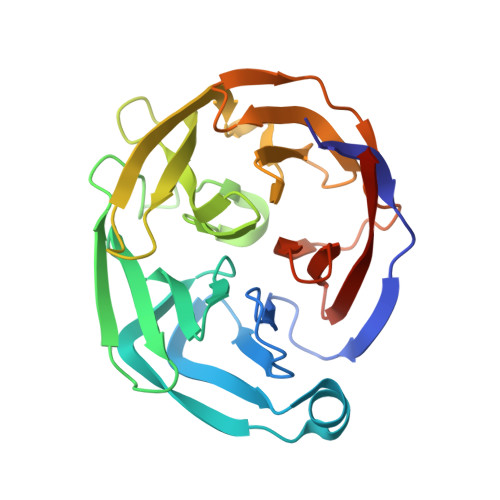 | |
UniProt & NIH Common Fund Data Resources | |||||
PHAROS: Q6ZMI3 GTEx: ENSG00000186417 | |||||
Entity Groups | |||||
| Sequence Clusters | 30% Identity50% Identity70% Identity90% Identity95% Identity100% Identity | ||||
| UniProt Group | Q6ZMI3 | ||||
Glycosylation | |||||
| Glycosylation Sites: 3 | Go to GlyGen: Q6ZMI3-1 | ||||
Sequence AnnotationsExpand | |||||
Reference Sequence | |||||
| Ligands 5 Unique | |||||
|---|---|---|---|---|---|
| ID | Chains | Name / Formula / InChI Key | 2D Diagram | 3D Interactions | |
| NAG Download:Ideal Coordinates CCD File | F [auth A], G [auth A], H [auth A] | 2-acetamido-2-deoxy-beta-D-glucopyranose C8 H15 N O6 OVRNDRQMDRJTHS-FMDGEEDCSA-N |  | ||
| PO4 Download:Ideal Coordinates CCD File | I [auth A] | PHOSPHATE ION O4 P NBIIXXVUZAFLBC-UHFFFAOYSA-K |  | ||
| EDO Download:Ideal Coordinates CCD File | J [auth A], K [auth A] | 1,2-ETHANEDIOL C2 H6 O2 LYCAIKOWRPUZTN-UHFFFAOYSA-N |  | ||
| CA Download:Ideal Coordinates CCD File | L [auth A], M [auth A] | CALCIUM ION Ca BHPQYMZQTOCNFJ-UHFFFAOYSA-N |  | ||
| NA Download:Ideal Coordinates CCD File | B [auth A], C [auth A], D [auth A], E [auth A] | SODIUM ION Na FKNQFGJONOIPTF-UHFFFAOYSA-N |  | ||
| Length ( Å ) | Angle ( ˚ ) |
|---|---|
| a = 46.88 | α = 90 |
| b = 81.573 | β = 90 |
| c = 66.194 | γ = 90 |
| Software Name | Purpose |
|---|---|
| PHENIX | refinement |
| HKL-3000 | phasing |


