5YAZ

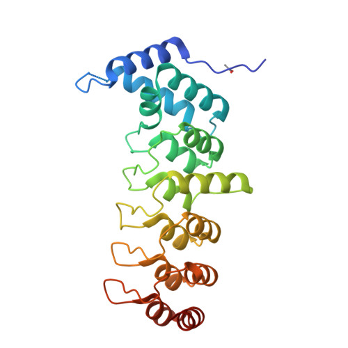
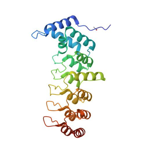
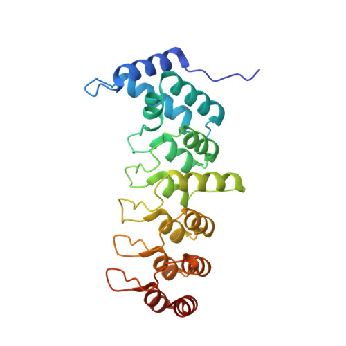
Crystal structure of the ANKRD domain of KANK1


Experimental Data Snapshot

  • Method: X-RAY DIFFRACTION
  • Resolution: 1.90 Å
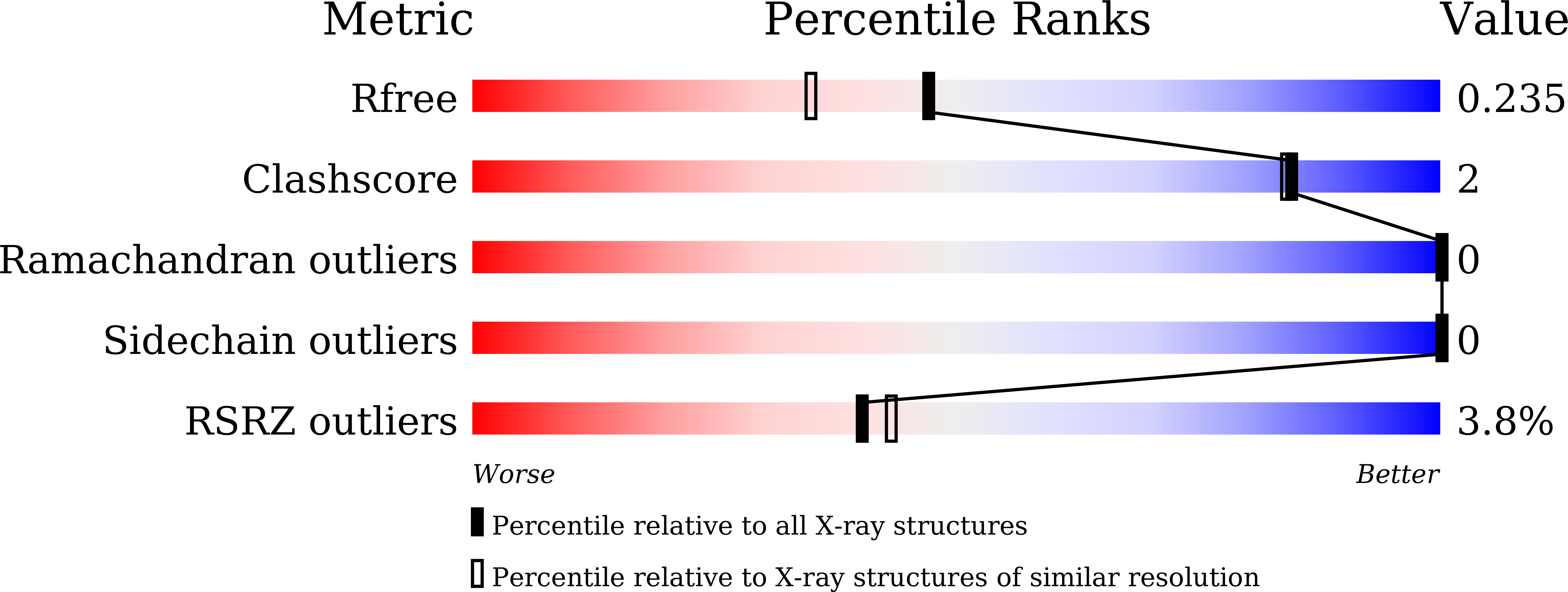
  • R-Value Free: 0.235 
  • R-Value Work: 0.193 
  • R-Value Observed: 0.196 

Starting Model: experimental
View more details

wwPDB Validation   3D Report Full Report


This is version 1.3 of the entry. See complete history


Literature

Structural insights into ankyrin repeat-mediated recognition of the kinesin motor protein KIF21A by KANK1, a scaffold protein in focal adhesion.

Pan, W.Sun, K.Tang, K.Xiao, Q.Ma, C.Yu, C.Wei, Z.

(2018) J Biol Chem 293: 1944-1956

  • DOI: https://doi.org/10.1074/jbc.M117.815779
  • Primary Citation of Related Structures:  
    5YAY, 5YAZ

  • PubMed Abstract: 

    Kidney ankyrin repeat-containing proteins (KANK1/2/3/4) belong to a family of scaffold proteins, playing critical roles in cytoskeleton organization, cell polarity, and migration. Mutations in KANK proteins are implicated in cancers and genetic diseases, such as nephrotic syndrome. KANK proteins can bind various target proteins through different protein regions, including a highly conserved ankyrin repeat domain (ANKRD). However, the molecular basis for target recognition by the ANKRD remains elusive. In this study, we solved a high-resolution crystal structure of the ANKRD of KANK1 in complex with a short sequence of the motor protein kinesin family member 21A (KIF21A), revealing that the highly specific target-binding mode of the ANKRD involves combinatorial use of two interfaces. Mutations in either interface disrupted the KANK1-KIF21A interaction. Cellular immunofluorescence localization analysis indicated that binding-deficient mutations block recruitment of KIF21A to focal adhesions by KANK1. In conclusion, our structural study provides mechanistic explanations for the ANKRD-mediated recognition of KIF21A and for many disease-related mutations identified in human KANK proteins.


  • Organizational Affiliation

    From the Department of Biology, Southern University of Science and Technology, 518055 Shenzhen, China.


Macromolecules
Find similar proteins by:  (by identity cutoff)  |  3D Structure
Entity ID: 1
MoleculeChains Sequence LengthOrganismDetailsImage
KN motif and ankyrin repeat domains 1257Mus musculusMutation(s): 0 
Gene Names: Kank1
UniProt & NIH Common Fund Data Resources
Find proteins for E9Q238 (Mus musculus)
Explore E9Q238 
Go to UniProtKB:  E9Q238
IMPC:  MGI:2147707
Entity Groups  
Sequence Clusters30% Identity50% Identity70% Identity90% Identity95% Identity100% Identity
UniProt GroupE9Q238
Sequence Annotations
Expand
  • Reference Sequence
Small Molecules
Ligands 1 Unique
IDChains Name / Formula / InChI Key2D Diagram3D Interactions
ACT
Query on ACT

Download Ideal Coordinates CCD File 
B [auth A]ACETATE ION
C2 H3 O2
QTBSBXVTEAMEQO-UHFFFAOYSA-M
Experimental Data & Validation

Experimental Data

  • Method: X-RAY DIFFRACTION
  • Resolution: 1.90 Å
  • R-Value Free: 0.235 
  • R-Value Work: 0.193 
  • R-Value Observed: 0.196 
  • Space Group: P 1 21 1
Unit Cell:
Length ( Å )Angle ( ˚ )
a = 29.102α = 90
b = 70.207β = 96.91
c = 60.655γ = 90
Software Package:
Software NamePurpose
HKL-2000data collection
HKL-2000data scaling
PHASERphasing
PHENIXrefinement
PDB_EXTRACTdata extraction

Structure Validation

View Full Validation Report


Entry History 

Deposition Data

Revision History  (Full details and data files)

  • Version 1.0: 2017-12-20
    Type: Initial release
  • Version 1.1: 2017-12-27
    Changes: Database references
  • Version 1.2: 2018-03-07
    Changes: Database references
  • Version 1.3: 2023-11-22
    Changes: Advisory, Data collection, Database references, Refinement description