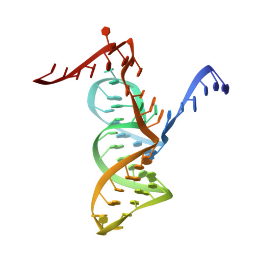
5Y87

Structure-based Insights into Self-Cleavage by a Four-way Junctional Twister-Sister Ribozyme


Experimental Data Snapshot

  • Method: X-RAY DIFFRACTION
  • Resolution: 2.13 Å
  • R-Value Free: 0.257 
  • R-Value Work: 0.202 
  • R-Value Observed: 0.205 

wwPDB Validation   3D Report Full Report


This is version 2.1 of the entry. See complete history


Literature

Structure-based insights into self-cleavage by a four-way junctional twister-sister ribozyme

Zheng, L.Mairhofer, E.Teplova, M.Zhang, Y.Ma, J.Patel, D.J.Micura, R.Ren, A.

(2017) Nat Commun 8: 1180-1180

  • DOI: https://doi.org/10.1038/s41467-017-01276-y
  • Primary Citation of Related Structures:  
    5Y85, 5Y87

  • PubMed Abstract: 

    Here we report on the crystal structure and cleavage assays of a four-way junctional twister-sister self-cleaving ribozyme. Notably, 11 conserved spatially separated loop nucleotides are brought into close proximity at the ribozyme core through long-range interactions mediated by hydrated Mg 2+ cations. The C62-A63 step at the cleavage site adopts a splayed-apart orientation, with flexible C62 directed outwards, whereas A63 is directed inwards and anchored by stacking and hydrogen-bonding interactions. Structure-guided studies of key base, sugar, and phosphate mutations in the twister-sister ribozyme, suggest contributions to the cleavage chemistry from interactions between a guanine at the active site and the non-bridging oxygen of the scissile phosphate, a feature found previously also for the related twister ribozyme. Our four-way junctional pre-catalytic structure differs significantly in the alignment at the cleavage step (splayed-apart vs. base-stacked) and surrounding residues and hydrated Mg 2+ ions relative to a reported three-way junctional pre-catalytic structure of the twister-sister ribozyme.


  • Organizational Affiliation

    Life Sciences Institute, Zhejiang University, 310058, Hangzhou, China.


Macromolecules

Find similar nucleic acids by:  Sequence   |   3D Structure  

Entity ID: 1
MoleculeChains LengthOrganismImage
RNA (5'-R(P*AP*CP*CP*CP*GP*CP*AP*AP*GP*GP*CP*CP*GP*AP*CP*GP*GP*C)-3')
A, C
18metagenome
Sequence Annotations
Expand
  • Reference Sequence
Find similar nucleic acids by: 3D Structure
Entity ID: 2
MoleculeChains LengthOrganismImage
DNA/RNA (50-MER)
B, D
50metagenome
Sequence Annotations
Expand
  • Reference Sequence
Small Molecules
Ligands 1 Unique
IDChains Name / Formula / InChI Key2D Diagram3D Interactions
MN
Query on MN

Download Ideal Coordinates CCD File 
E [auth A]
F [auth A]
G [auth B]
H [auth B]
I [auth B]
E [auth A],
F [auth A],
G [auth B],
H [auth B],
I [auth B],
J [auth B],
K [auth B],
L [auth B],
M [auth B],
N [auth B],
O [auth B],
P [auth B],
Q [auth B],
R [auth C],
S [auth D],
T [auth D],
U [auth D],
V [auth D],
W [auth D],
X [auth D],
Y [auth D],
Z [auth D]
MANGANESE (II) ION
Mn
WAEMQWOKJMHJLA-UHFFFAOYSA-N
Experimental Data & Validation

Experimental Data

  • Method: X-RAY DIFFRACTION
  • Resolution: 2.13 Å
  • R-Value Free: 0.257 
  • R-Value Work: 0.202 
  • R-Value Observed: 0.205 
  • Space Group: P 21 21 2
Unit Cell:
Length ( Å )Angle ( ˚ )
a = 91.221α = 90
b = 108.963β = 90
c = 41.794γ = 90
Software Package:
Software NamePurpose
PHENIXrefinement
HKL-2000data reduction
HKL-2000data scaling
PHASERphasing

Structure Validation

View Full Validation Report


Entry History 

Deposition Data

Revision History  (Full details and data files)

  • Version 1.0: 2017-11-22
    Type: Initial release
  • Version 2.0: 2019-01-23
    Changes: Atomic model, Data collection, Derived calculations
  • Version 2.1: 2024-03-27
    Changes: Data collection, Database references, Derived calculations