


Crystal structure of the bile salt hydrolase from lactobacillus salivarius complex with glycocholic acid and cholic acid
Xu, F., Hu, X.-J., Lin, J.To be published.
Experimental Data Snapshot
Starting Model: experimental
View more details
Entity ID: 1 | |||||
|---|---|---|---|---|---|
| Molecule | Chains | Sequence Length | Organism | Details | Image |
| Bile salt hydrolase | 333 | Ligilactobacillus salivarius | Mutation(s): 0 EC: 3.5.1.24 | 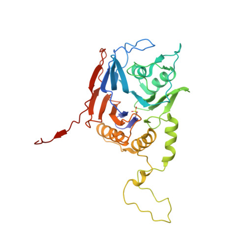 | |
UniProt | |||||
Entity Groups | |||||
| Sequence Clusters | 30% Identity50% Identity70% Identity90% Identity95% Identity100% Identity | ||||
| UniProt Group | J7H3P9 | ||||
Sequence AnnotationsExpand | |||||
Reference Sequence | |||||
| Ligands 6 Unique | |||||
|---|---|---|---|---|---|
| ID | Chains | Name / Formula / InChI Key | 2D Diagram | 3D Interactions | |
| GCH Download:Ideal Coordinates CCD File | I [auth A], J [auth A], S [auth F] | GLYCOCHOLIC ACID C26 H43 N O6 RFDAIACWWDREDC-FRVQLJSFSA-N |  | ||
| CHD Download:Ideal Coordinates CCD File | K [auth B] N [auth C] O [auth D] Q [auth E] U [auth G] | CHOLIC ACID C24 H40 O5 BHQCQFFYRZLCQQ-OELDTZBJSA-N |  | ||
| PEG Download:Ideal Coordinates CCD File | T [auth F] | DI(HYDROXYETHYL)ETHER C4 H10 O3 MTHSVFCYNBDYFN-UHFFFAOYSA-N |  | ||
| PO4 Download:Ideal Coordinates CCD File | M [auth B] | PHOSPHATE ION O4 P NBIIXXVUZAFLBC-UHFFFAOYSA-K |  | ||
| GOL Download:Ideal Coordinates CCD File | L [auth B], R [auth E] | GLYCEROL C3 H8 O3 PEDCQBHIVMGVHV-UHFFFAOYSA-N |  | ||
| EDO Download:Ideal Coordinates CCD File | P [auth D], V [auth G], X [auth H] | 1,2-ETHANEDIOL C2 H6 O2 LYCAIKOWRPUZTN-UHFFFAOYSA-N |  | ||
| Modified Residues 1 Unique | |||||
|---|---|---|---|---|---|
| ID | Chains | Type | Formula | 2D Diagram | Parent |
| OCS Query on OCS | A, B, C, D, E A, B, C, D, E, F, G, H | L-PEPTIDE LINKING | C3 H7 N O5 S |  | CYS |
| Length ( Å ) | Angle ( ˚ ) |
|---|---|
| a = 84.01 | α = 90 |
| b = 94.09 | β = 90.64 |
| c = 166.974 | γ = 90 |
| Software Name | Purpose |
|---|---|
| REFMAC | refinement |
| iMOSFLM | data reduction |
| Aimless | data scaling |
| PHASER | phasing |


