5Y4H | pdb_00005y4h

Human SIRT3 in complex with halistanol sulfate


Experimental Data Snapshot

  • Method: X-RAY DIFFRACTION
  • Resolution: 2.60 Å
  • R-Value Free: 
    0.269 (Depositor), 0.270 (DCC) 
  • R-Value Work: 
    0.215 (Depositor), 0.220 (DCC) 
  • R-Value Observed: 
    0.218 (Depositor) 

Starting Model: experimental
View more details

wwPDB Validation   3D Report Full Report


Ligand Structure Quality Assessment 

Created with Raphaël 2.3.0Worse 01 BetterLigand structure goodness of fit to experimental dataBest fitted 8QFClick on this verticalbar to view details

This is version 1.2 of the entry. See complete history


Literature

Halistanol sulfates I and J, new SIRT1-3 inhibitory steroid sulfates from a marine sponge of the genus Halichondria

Nakamura, F.Kudo, N.Tomachi, Y.Nakata, A.Takemoto, M.Ito, A.Tabei, H.Arai, D.de Voogd, N.Yoshida, M.Nakao, Y.Fusetani, N.

(2018) J Antibiot (Tokyo) 71: 273-278

  • DOI: https://doi.org/10.1038/ja.2017.145
  • Primary Citation of Related Structures:  
    5Y4H

  • PubMed Abstract: 

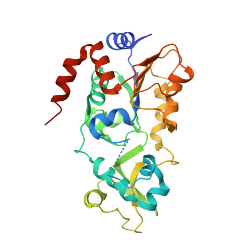
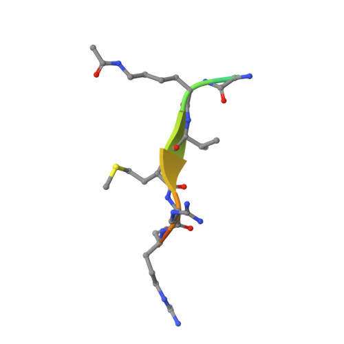
    Two new analogs of halistanol sulfate (1) were isolated from a marine sponge Halichondria sp. collected at Hachijo-jima Island. Structures of these new halistanol sulfates I (2) and J (3) were elucidated by spectral analyses. Compounds 1-3 showed inhibitory activity against SIRT 1-3 with IC 50 ranges of 45.9-67.9, 18.9-21.1 and 21.8-37.5 μM, respectively. X-ray crystallography of the halistanol sulfate (1) and SIRT3 complex clearly indicates that 1 binds to the exosite of SIRT3 that we have discovered in this study.


  • Organizational Affiliation

    Department of Chemistry and Biochemistry, Graduate School of Advanced Science and Engineering, Waseda University, Tokyo, Japan.


Macromolecules
Find similar proteins by:  (by identity cutoff)  |  3D Structure
Entity ID: 1
MoleculeChains Sequence LengthOrganismDetailsImage
NAD-dependent protein deacetylase sirtuin-3, mitochondrial285Homo sapiensMutation(s): 0 
Gene Names: SIRT3SIR2L3
EC: 3.5.1 (PDB Primary Data), 2.3.1 (UniProt), 2.3.1.286 (UniProt)
UniProt & NIH Common Fund Data Resources
Find proteins for Q9NTG7 (Homo sapiens)
Explore Q9NTG7 
Go to UniProtKB:  Q9NTG7
PHAROS:  Q9NTG7
GTEx:  ENSG00000142082 
Entity Groups  
Sequence Clusters30% Identity50% Identity70% Identity90% Identity95% Identity100% Identity
UniProt GroupQ9NTG7
Sequence Annotations
Expand
  • Reference Sequence

Find similar proteins by:  Sequence   |   3D Structure  

Entity ID: 2
MoleculeChains Sequence LengthOrganismDetailsImage
THr-Arg-Ser-GLY-ALY-VAL-MET-ARG-ARG-LEU-ARG-ARG12Homo sapiensMutation(s): 0 
EC: 6.2.1.1 (UniProt), 6.2.1.17 (UniProt)
UniProt & NIH Common Fund Data Resources
Find proteins for Q9NUB1 (Homo sapiens)
Explore Q9NUB1 
Go to UniProtKB:  Q9NUB1
PHAROS:  Q9NUB1
GTEx:  ENSG00000154930 
Entity Groups  
Sequence Clusters30% Identity50% Identity70% Identity90% Identity95% Identity100% Identity
UniProt GroupQ9NUB1
Sequence Annotations
Expand
  • Reference Sequence
Small Molecules
Ligands 2 Unique
IDChains Name / Formula / InChI Key2D Diagram3D Interactions
8QF
Query on 8QF

Download Ideal Coordinates CCD File 
D [auth A][(2~{S},3~{S},5~{S},6~{S},8~{S},9~{S},10~{R},13~{R},14~{R},17~{R})-17-[(2~{R})-6,6-dimethylheptan-2-yl]-10,13-dimethyl-2,3-disulfooxy-2,3,4,5,6,7,8,9,11,12,14,15,16,17-tetradecahydro-1~{H}-cyclopenta[a]phenanthren-6-yl] hydrogen sulfate
C28 H50 O12 S3
MNVNGGMQJFYHMK-NZLCMVQPSA-N
ZN
Query on ZN

Download Ideal Coordinates CCD File 
C [auth A]ZINC ION
Zn
PTFCDOFLOPIGGS-UHFFFAOYSA-N
Modified Residues  1 Unique
IDChains TypeFormula2D DiagramParent
ALY
Query on ALY
B
L-PEPTIDE LINKINGC8 H16 N2 O3LYS
Experimental Data & Validation

Experimental Data

  • Method: X-RAY DIFFRACTION
  • Resolution: 2.60 Å
  • R-Value Free:  0.269 (Depositor), 0.270 (DCC) 
  • R-Value Work:  0.215 (Depositor), 0.220 (DCC) 
  • R-Value Observed: 0.218 (Depositor) 
Space Group: P 4 3 2
Unit Cell:
Length ( Å )Angle ( ˚ )
a = 127.84α = 90
b = 127.84β = 90
c = 127.84γ = 90
Software Package:
Software NamePurpose
REFMACrefinement
HKL-2000data reduction
HKL-2000data scaling
MOLREPphasing

Structure Validation

View Full Validation Report


Ligand Structure Quality Assessment 

Created with Raphaël 2.3.0Worse 01 BetterLigand structure goodness of fit to experimental dataBest fitted 8QFClick on this verticalbar to view details

Entry History & Funding Information

Deposition Data


Funding OrganizationLocationGrant Number
JSPSJapanKAKENHI 26221204
JSPSJapanP-Direct

Revision History  (Full details and data files)

  • Version 1.0: 2018-05-16
    Type: Initial release
  • Version 1.1: 2023-11-22
    Changes: Data collection, Database references, Derived calculations, Refinement description
  • Version 1.2: 2024-10-23
    Changes: Structure summary