5Y3N | pdb_00005y3n

Structure of TRAP1 complexed with DN401


Experimental Data Snapshot

  • Method: X-RAY DIFFRACTION
  • Resolution: 2.40 Å
  • R-Value Free: 
    0.257 (Depositor), 0.266 (DCC) 
  • R-Value Work: 
    0.208 (Depositor) 
  • R-Value Observed: 
    0.210 (Depositor) 

wwPDB Validation 3D Report Full Report

Validation slider image for 5Y3N

Ligand Structure Quality Assessment 


This is version 1.2 of the entry. See complete history

Literature

Paralog Specificity Determines Subcellular Distribution, Action Mechanism, and Anticancer Activity of TRAP1 Inhibitors.

Park, H.K.Jeong, H.Ko, E.Lee, G.Lee, J.E.Lee, S.K.Lee, A.J.Im, J.Y.Hu, S.Kim, S.H.Lee, J.H.Lee, C.Kang, S.Kang, B.H.

(2017) J Med Chem 60: 7569-7578

  • DOI: https://doi.org/10.1021/acs.jmedchem.7b00978
  • Primary Citation Related Structures: 
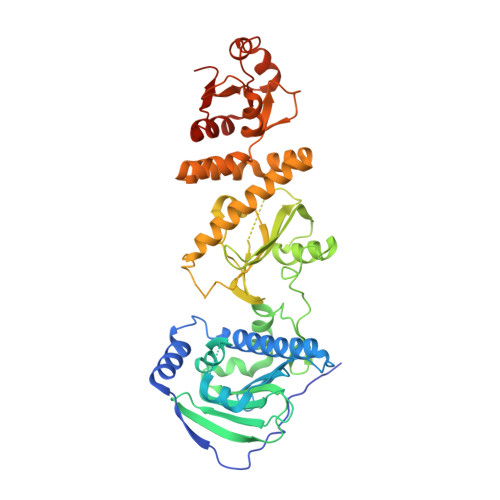
    5Y3N, 5Y3O

  • PubMed Abstract: 

    Although Hsp90 inhibitors can inhibit multiple tumorigenic pathways in cancer cells, their anticancer activity has been disappointingly modest. However, by forcing Hsp90 inhibitors into the mitochondria with mitochondrial delivery vehicles, they were converted into potent drugs targeting the mitochondrial Hsp90 paralog TRAP1. Here, to improve mitochondrial drug accumulation without using the mitochondrial delivery vehicle, we increased freely available drug concentrations in the cytoplasm by reducing the binding of the drugs to the abundant cytoplasmic Hsp90. After analyzing X-ray cocrystal structures, the purine ring of the Hsp90 inhibitor 2 (BIIB021) was modified to pyrazolopyrimidine scaffolds. One pyrazolopyrimidine, 12b (DN401), bound better to TRAP1 than to Hsp90, inactivated the mitochondrial TRAP1 in vivo, and it exhibited potent anticancer activity. Therefore, the rationale and feasible guidelines for developing 12b can potentially be exploited to design a potent TRAP1 inhibitor.


  • Organizational Affiliation
    • Department of Biological Sciences, Ulsan National Institutes of Science and Technology (UNIST) , Ulsan 44919, South Korea.

Macromolecule Content 

  • Total Structure Weight: 57.8 kDa 
  • Atom Count: 3,630 
  • Modeled Residue Count: 438 
  • Deposited Residue Count: 502 
  • Unique protein chains: 1

Macromolecules

Find similar proteins by:|  3D Structure
Entity ID: 1
MoleculeChains  Sequence LengthOrganismDetailsImage
Heat shock protein 75 kDa, mitochondrial502Homo sapiensMutation(s): 0 
Gene Names: TRAP1HSP75
UniProt & NIH Common Fund Data Resources
Find proteins for Q12931 (Homo sapiens)
Explore Q12931 
Go to UniProtKB:  Q12931
PHAROS:  Q12931
GTEx:  ENSG00000126602 
Entity Groups
Sequence Clusters30% Identity50% Identity70% Identity90% Identity95% Identity100% Identity
UniProt GroupQ12931
Sequence Annotations
Expand
Reference Sequence

Small Molecules

Ligands 1 Unique
IDChains Name / Formula / InChI Key2D Diagram3D Interactions
8MF

Query on 8MF


Download:Ideal Coordinates CCD File
B [auth A]1-[(6-bromanyl-1,3-benzodioxol-5-yl)methyl]-4-chloranyl-pyrazolo[3,4-d]pyrimidin-6-amine
C13 H9 Br Cl N5 O2
BIABSXPPEYWVER-UHFFFAOYSA-N
Binding Affinity Annotations 
IDSourceBinding Affinity
8MF BindingDB:  5Y3N IC50: min: 79, max: 143 (nM) from 2 assay(s)

Experimental Data & Validation

Experimental Data

  • Method: X-RAY DIFFRACTION
  • Resolution: 2.40 Å
  • R-Value Free:  0.257 (Depositor), 0.266 (DCC) 
  • R-Value Work:  0.208 (Depositor) 
  • R-Value Observed: 0.210 (Depositor) 
Space Group: P 41 21 2
Unit Cell:
Length ( Å )Angle ( ˚ )
a = 69.41α = 90
b = 69.41β = 90
c = 252.159γ = 90
Software Package:
Software NamePurpose
PHENIXrefinement
HKL-2000data reduction
HKL-2000data scaling
PHENIXphasing
Cootmodel building

Structure Validation

View Full Validation Report


Ligand Structure Quality Assessment 


Entry History 

Deposition Data

Revision History  (Full details and data files)

  • Version 1.0: 2017-08-30
    Type: Initial release
  • Version 1.1: 2017-12-06
    Changes: Database references
  • Version 1.2: 2024-03-27
    Changes: Data collection, Database references