Structural basis of the correct subunit assembly, aggregation, and intracellular degradation of nylon hydrolase
Negoro, S., Shibata, N., Lee, Y.H., Takehara, I., Kinugasa, R., Nagai, K., Tanaka, Y., Kato, D.I., Takeo, M., Goto, Y., Higuchi, Y.(2018) Sci Rep 8: 9725-9725
- PubMed: 29950566
- DOI: https://doi.org/10.1038/s41598-018-27860-w
- Primary Citation of Related Structures:
5XYG, 5XYO, 5XYP, 5XYQ, 5XYS, 5XYT, 5Y0L, 5Y0M - PubMed Abstract:
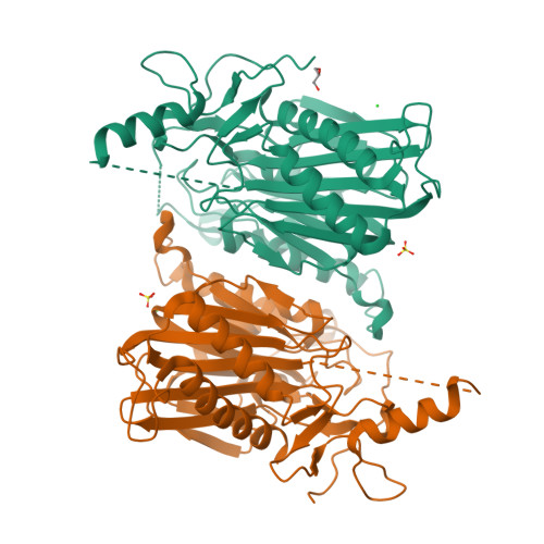
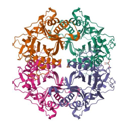
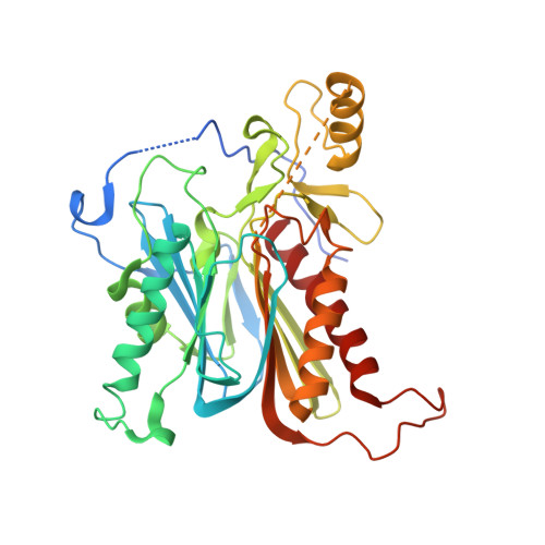
Nylon hydrolase (NylC) is initially expressed as an inactive precursor (36 kDa). The precursor is cleaved autocatalytically at Asn266/Thr267 to generate an active enzyme composed of an α subunit (27 kDa) and a β subunit (9 kDa). Four αβ heterodimers (molecules A-D) form a doughnut-shaped quaternary structure. In this study, the thermostability of the parental NylC was altered by amino acid substitutions located at the A/D interface (D122G/H130Y/D36A/L137A) or the A/B interface (E263Q) and spanned a range of 47 °C. Considering structural, biophysical, and biochemical analyses, we discuss the structural basis of the stability of nylon hydrolase. From the analytical centrifugation data obtained regarding the various mutant enzymes, we conclude that the assembly of the monomeric units is dynamically altered by the mutations. Finally, we propose a model that can predict whether the fate of the nascent polypeptide will be correct subunit assembly, inappropriate protein-protein interactions causing aggregation, or intracellular degradation of the polypeptide.
Organizational Affiliation:
Department of Applied Chemistry, Graduate School of Engineering, University of Hyogo, Himeji, 671-2280, Japan. negoro@eng.u-hyogo.ac.jp.


