5XXM | pdb_00005xxm

Crystal structure of GH3 beta-glucosidase from Bacteroides thetaiotaomicron in complex with gluconolactone


Experimental Data Snapshot

  • Method: X-RAY DIFFRACTION
  • Resolution: 1.70 Å
  • R-Value Free: 
    0.208 (Depositor), 0.215 (DCC) 
  • R-Value Work: 
    0.181 (Depositor), 0.190 (DCC) 
  • R-Value Observed: 
    0.183 (Depositor) 

wwPDB Validation 3D Report Full Report

Validation slider image for 5XXM

Ligand Structure Quality Assessment 


This is version 1.3 of the entry. See complete history

Literature

Function and structure relationships of a beta-1,2-glucooligosaccharide-degrading beta-glucosidase.

Ishiguro, R.Tanaka, N.Abe, K.Nakajima, M.Maeda, T.Miyanaga, A.Takahashi, Y.Sugimoto, N.Nakai, H.Taguchi, H.

(2017) FEBS Lett 591: 3926-3936

  • DOI: https://doi.org/10.1002/1873-3468.12911
  • Primary Citation Related Structures: 
    5XXL, 5XXM, 5XXN, 5XXO

  • PubMed Abstract: 

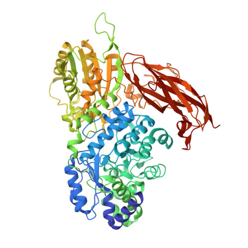
    BT_3567 protein, a putative β-glucosidase from Bacteroides thetaiotaomicron, exhibits higher activity toward Sop 3-5 (Sop n , n: degree of polymerization of β-1,2-glucooligosaccharides) than toward Sop 2 , unlike a known β-glucosidase from Listeria innocua which predominantly prefers Sop 2 . In the complex structure determined by soaking of a D286N mutant crystal with Sop 4 , a Sop 3 moiety was observed at subsites -1 to +2. The glucose moiety at subsite +2 forms a hydrogen bond with Asn81, which is replaced with Gly in the L. innocua β-glucosidase. The K m values of the N81G mutant for Sop 3-5 are much higher than those of the wild-type, suggesting that Asn81 contributes to the binding to substrates longer than Sop 3 .


  • Organizational Affiliation
    • Department of Applied Biological Science, Faculty of Science and Technology, Tokyo University of Science, Noda, Chiba, Japan.

Macromolecule Content 

  • Total Structure Weight: 168.64 kDa 
  • Atom Count: 12,778 
  • Modeled Residue Count: 1,493 
  • Deposited Residue Count: 1,520 
  • Unique protein chains: 1

Macromolecules

Find similar proteins by:|  3D Structure
Entity ID: 1
MoleculeChains  Sequence LengthOrganismDetailsImage
Periplasmic beta-glucosidase
A, B
760Bacteroides thetaiotaomicron VPI-5482Mutation(s): 0 
Gene Names: BT_3567
EC: 3.2.1.21
UniProt
Find proteins for Q8A1U1 (Bacteroides thetaiotaomicron (strain ATCC 29148 / DSM 2079 / JCM 5827 / CCUG 10774 / NCTC 10582 / VPI-5482 / E50))
Explore Q8A1U1 
Go to UniProtKB:  Q8A1U1
Entity Groups
Sequence Clusters30% Identity50% Identity70% Identity90% Identity95% Identity100% Identity
UniProt GroupQ8A1U1
Sequence Annotations
Expand
Reference Sequence

Experimental Data & Validation

Experimental Data

  • Method: X-RAY DIFFRACTION
  • Resolution: 1.70 Å
  • R-Value Free:  0.208 (Depositor), 0.215 (DCC) 
  • R-Value Work:  0.181 (Depositor), 0.190 (DCC) 
  • R-Value Observed: 0.183 (Depositor) 
Space Group: C 2 2 21
Unit Cell:
Length ( Å )Angle ( ˚ )
a = 81.82α = 90
b = 167.69β = 90
c = 224.9γ = 90
Software Package:
Software NamePurpose
REFMACrefinement
iMOSFLMdata reduction
MOLREPphasing

Structure Validation

View Full Validation Report


Ligand Structure Quality Assessment 


Entry History 

Deposition Data

Revision History  (Full details and data files)

  • Version 1.0: 2017-12-13
    Type: Initial release
  • Version 1.1: 2017-12-27
    Changes: Database references
  • Version 1.2: 2020-07-29
    Type: Remediation
    Reason: Carbohydrate remediation
    Changes: Derived calculations, Structure summary
  • Version 1.3: 2024-03-27
    Changes: Data collection, Database references, Structure summary