5XWF | pdb_00005xwf

Crystal structure of chitinase (RmChi1) from Rhizomucor miehei (SP3221/SAD)


Experimental Data Snapshot

  • Method: X-RAY DIFFRACTION
  • Resolution: 2.58 Å
  • R-Value Free: 
    0.206 (Depositor), 0.211 (DCC) 
  • R-Value Work: 
    0.172 (Depositor), 0.175 (DCC) 
  • R-Value Observed: 
    0.175 (Depositor) 

wwPDB Validation 3D Report Full Report

Validation slider image for 5XWF

This is version 1.2 of the entry. See complete history

Literature

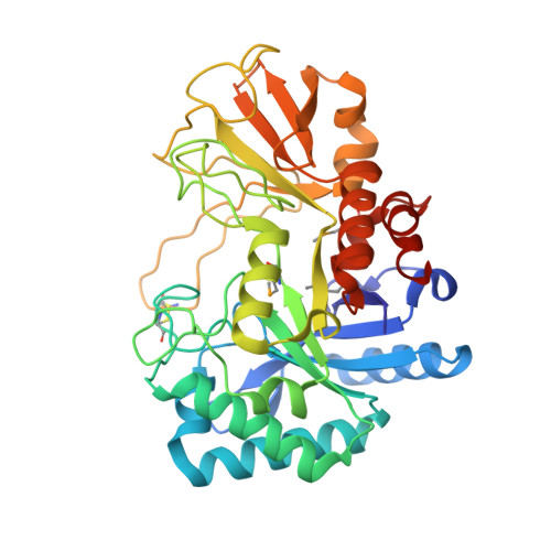
Crystal structure of a chitinase (RmChiA) from the thermophilic fungus Rhizomucor miehei with a real active site tunnel.

Jiang, Z.Q.Hu, S.Q.Ma, J.Liu, Y.C.Qiao, Z.Yan, Q.J.Gao, Y.Yang, S.Q.

(2021) Biochim Biophys Acta Proteins Proteom : 140709-140709

  • DOI: https://doi.org/10.1016/j.bbapap.2021.140709
  • Primary Citation Related Structures: 
    5XWF, 5YUQ, 7FBT

  • PubMed Abstract: 

    A chitinase gene (RmChiA) encoding 445 amino acid (aa) residues from a fungus Rhizomucor miehei was cloned and overexpressed in Escherichia coli. Two kinds of RmChiA crystal forms, with space groups P3 2 2 1 and P1, were obtained by sitting-drop vapor diffusion and the structures were determined by X-ray diffraction. The overall structure of RmChiA monomer, which is the first structure of bacterial-type chitinases from nonpathogenic fungi, adopts a canonical triosephosphate isomerase (TIM) barrel fold with two protruding chitinase insertion domains. RmChiA exhibited a unique NxDxE catalytical motif and a real active site tunnel structure, which are firstly found in GH family 18 chitinases. The motif had high structural homolog with the typical DxDxE motif in other GH family 18 chitinases. The tunnel is formed by two unusual long loops, containing 15 aa and 45 aa respectively, linked by a disulfide bond across the substrate-binding cleft. Mutation experiments found that opening the roof of tunnel structure increased the hydrolysis efficiency of RmChiA, but the thermostability of the mutants decreased. Moreover, the tunnel structure endowed RmChiA with the exo-chitinase character.


  • Organizational Affiliation
    • College of Food Science and Nutritional Engineering, China Agricultural University, Beijing 100083, China.

Macromolecule Content 

  • Total Structure Weight: 41.6 kDa 
  • Atom Count: 2,957 
  • Modeled Residue Count: 369 
  • Deposited Residue Count: 378 
  • Unique protein chains: 1

Macromolecules

Find similar proteins by:|  3D Structure
Entity ID: 1
MoleculeChains  Sequence LengthOrganismDetailsImage
Fungal chitinase from Rhizomucor miehei (SeMet-substituted proteins)378Rhizomucor mieheiMutation(s): 0 
EC: 3.2.1.14
UniProt
Find proteins for A0A3B6UEQ2 (Rhizomucor miehei)
Explore A0A3B6UEQ2 
Go to UniProtKB:  A0A3B6UEQ2
Entity Groups
Sequence Clusters30% Identity50% Identity70% Identity90% Identity95% Identity100% Identity
UniProt GroupA0A3B6UEQ2
Sequence Annotations
Expand
Reference Sequence

Small Molecules

Modified Residues  1 Unique
IDChains TypeFormula2D DiagramParent
MSE
Query on MSE
A
L-PEPTIDE LINKINGC5 H11 N O2 SeMET

Experimental Data & Validation

Experimental Data

  • Method: X-RAY DIFFRACTION
  • Resolution: 2.58 Å
  • R-Value Free:  0.206 (Depositor), 0.211 (DCC) 
  • R-Value Work:  0.172 (Depositor), 0.175 (DCC) 
  • R-Value Observed: 0.175 (Depositor) 
Space Group: P 32 2 1
Unit Cell:
Length ( Å )Angle ( ˚ )
a = 48.811α = 90
b = 48.811β = 90
c = 247.516γ = 120
Software Package:
Software NamePurpose
PHENIXrefinement
XDSdata reduction
XDSdata scaling
PHENIXphasing

Structure Validation

View Full Validation Report


Entry History 

& Funding Information

Deposition Data


Funding OrganizationLocationGrant Number
the National Science Fund for Distinguished Young ScholarsChina31325021

Revision History  (Full details and data files)

  • Version 1.0: 2018-07-04
    Type: Initial release
  • Version 1.1: 2021-08-18
    Changes: Database references
  • Version 1.2: 2024-10-30
    Changes: Data collection, Structure summary