5XVU | pdb_00005xvu

Crystal structure of the protein kinase CK2 catalytic domain from Plasmodium falciparum bound to ATP


Experimental Data Snapshot

  • Method: X-RAY DIFFRACTION
  • Resolution: 3.00 Å
  • R-Value Free: 
    0.216 (Depositor), 0.197 (DCC) 
  • R-Value Work: 
    0.172 (Depositor) 
  • R-Value Observed: 
    0.174 (Depositor) 

Starting Model: experimental
View more details

wwPDB Validation 3D Report Full Report

Validation slider image for 5XVU

Ligand Structure Quality Assessment 


This is version 1.3 of the entry. See complete history

Literature

The protein kinase CK2 catalytic domain from Plasmodium falciparum: crystal structure, tyrosine kinase activity and inhibition.

Ruiz-Carrillo, D.Lin, J.Q.El Sahili, A.Wei, M.Sze, S.K.Cheung, P.C.F.Doerig, C.Lescar, J.

(2018) Sci Rep 8: 7365-7365

  • DOI: https://doi.org/10.1038/s41598-018-25738-5
  • Primary Citation Related Structures: 
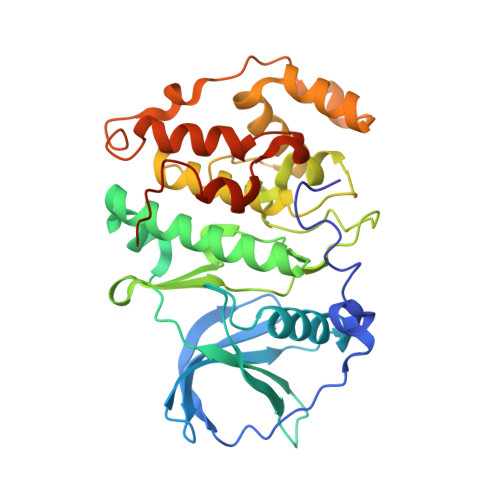
    5XVU

  • PubMed Abstract: 

    Malaria causes every year over half-a-million deaths. The emergence of parasites resistant to available treatments makes the identification of new targets and their inhibitors an urgent task for the development of novel anti-malaria drugs. Protein kinase CK2 is an evolutionary-conserved eukaryotic serine/threonine protein kinase that in Plasmodium falciparum (PfCK2) has been characterized as a promising target for chemotherapeutic intervention against malaria. Here we report a crystallographic structure of the catalytic domain of PfCK2α (D179S inactive single mutant) in complex with ATP at a resolution of 3.0 Å. Compared to the human enzyme, the structure reveals a subtly altered ATP binding pocket comprising five substitutions in the vicinity of the adenine base, that together with potential allosteric sites, could be exploited to design novel inhibitors specifically targeting the Plasmodium enzyme. We provide evidence for the dual autophosphorylation of residues Thr 63 and Tyr 30 of PfCK2. We also show that CX4945, a human CK2 inhibitor in clinical trials against solid tumor cancers, is effective against PfCK2 with an IC 50 of 13.2 nM.


  • Organizational Affiliation
    • School of Biological Sciences, 60 Nanyang Drive, Nanyang Technological University, Singapore, 637551, Singapore.

Macromolecule Content 

  • Total Structure Weight: 121.58 kDa 
  • Atom Count: 8,416 
  • Modeled Residue Count: 963 
  • Deposited Residue Count: 996 
  • Unique protein chains: 1

Macromolecules

Find similar proteins by:|  3D Structure
Entity ID: 1
MoleculeChains  Sequence LengthOrganismDetailsImage
Casein kinase 2, alpha subunit
A, B, C
332Plasmodium falciparum 3D7Mutation(s): 0 
Gene Names: PF11_0096PF3D7_1108400
EC: 2.7.11.1
UniProt
Find proteins for Q8IIR9 (Plasmodium falciparum (isolate 3D7))
Explore Q8IIR9 
Go to UniProtKB:  Q8IIR9
Entity Groups
Sequence Clusters30% Identity50% Identity70% Identity90% Identity95% Identity100% Identity
UniProt GroupQ8IIR9
Sequence Annotations
Expand
Reference Sequence

Small Molecules

Ligands 5 Unique
IDChains Name / Formula / InChI Key2D Diagram3D Interactions
ATP
(Subject of Investigation/LOI)

Query on ATP


Download:Ideal Coordinates CCD File
F [auth A],
L [auth B],
R [auth C]
ADENOSINE-5'-TRIPHOSPHATE
C10 H16 N5 O13 P3
ZKHQWZAMYRWXGA-KQYNXXCUSA-N
PEG

Query on PEG


Download:Ideal Coordinates CCD File
Q [auth C]DI(HYDROXYETHYL)ETHER
C4 H10 O3
MTHSVFCYNBDYFN-UHFFFAOYSA-N
SO4

Query on SO4


Download:Ideal Coordinates CCD File
D [auth A],
I [auth B],
J [auth B],
N [auth C],
O [auth C]
SULFATE ION
O4 S
QAOWNCQODCNURD-UHFFFAOYSA-L
EDO

Query on EDO


Download:Ideal Coordinates CCD File
G [auth A],
H [auth A],
M [auth B],
S [auth C]
1,2-ETHANEDIOL
C2 H6 O2
LYCAIKOWRPUZTN-UHFFFAOYSA-N
CL

Query on CL


Download:Ideal Coordinates CCD File
E [auth A],
K [auth B],
P [auth C]
CHLORIDE ION
Cl
VEXZGXHMUGYJMC-UHFFFAOYSA-M

Experimental Data & Validation

Experimental Data

  • Method: X-RAY DIFFRACTION
  • Resolution: 3.00 Å
  • R-Value Free:  0.216 (Depositor), 0.197 (DCC) 
  • R-Value Work:  0.172 (Depositor) 
  • R-Value Observed: 0.174 (Depositor) 
Space Group: P 21 21 21
Unit Cell:
Length ( Å )Angle ( ˚ )
a = 88.41α = 90
b = 123.55β = 90
c = 125.65γ = 90
Software Package:
Software NamePurpose
BUSTERrefinement
XDSdata reduction
XDSdata scaling
PHASERphasing

Structure Validation

View Full Validation Report


Ligand Structure Quality Assessment 


Entry History 

Deposition Data

Revision History  (Full details and data files)

  • Version 1.0: 2017-07-12
    Type: Initial release
  • Version 1.1: 2019-01-23
    Changes: Data collection, Database references
  • Version 1.2: 2021-11-03
    Changes: Database references, Structure summary
  • Version 1.3: 2023-11-22
    Changes: Data collection, Refinement description