5XTU | pdb_00005xtu

Crystal Structure of GDSL Esterase of Photobacterium sp. J15


Experimental Data Snapshot

  • Method: X-RAY DIFFRACTION
  • Resolution: 1.38 Å
  • R-Value Free: 
    0.179 (Depositor), 0.187 (DCC) 
  • R-Value Work: 
    0.158 (Depositor), 0.166 (DCC) 
  • R-Value Observed: 
    0.159 (Depositor) 

wwPDB Validation 3D Report Full Report

Validation slider image for 5XTU

Ligand Structure Quality Assessment 


This is version 1.2 of the entry. See complete history

Literature

Crystallization and structure elucidation of GDSL esterase of Photobacterium sp. J15.

Mazlan, S.N.H.S.Ali, M.S.M.Rahman, R.N.Z.R.A.Sabri, S.Jonet, M.A.Leow, T.C.

(2018) Int J Biol Macromol 119: 1188-1194

  • DOI: https://doi.org/10.1016/j.ijbiomac.2018.08.022
  • Primary Citation Related Structures: 
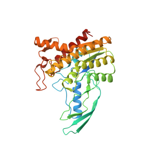
    5XTU

  • PubMed Abstract: 

    GDSL esterase J15 (EstJ15) is a member of Family II of lipolytic enzyme. The enzyme was further classified in subgroup SGNH hydrolase due to the presence of highly conserve motif, Ser-Gly-Asn-His in four conserved blocks I, II, III, and V, respectively. X-ray quality crystal of EstJ15 was obtained from optimized formulation containing 0.10 M ammonium sulphate, 0.15 M sodium cacodylate trihydrate pH 6.5, and 20% PEG 8000. The crystal structure of EstJ15 was solved at 1.38 Å with one molecule per asymmetric unit. The structure exhibits α/β hydrolase fold and shared low amino acid sequence identity of 23% with the passenger domain of the autotransporter EstA of Pseudomonas aeruginosa. The active site is located at the centre of the structure, formed a narrow tunnel that hinder long substrates to be catalysed which was proven by the protein-ligand docking analysis. This study facilitates the understanding of high substrate specificity of EstJ15 and provide insights on its catalytic mechanism.


  • Organizational Affiliation
    • Department of Cell and Molecular Biology, Faculty of Biotechnology and Biomolecular Sciences, Universiti Putra Malaysia, 43400 UPM Serdang, Selangor, Malaysia; Enzyme and Microbial Technology Research Center, Malaysia.

Macromolecule Content 

  • Total Structure Weight: 39.81 kDa 
  • Atom Count: 3,042 
  • Modeled Residue Count: 328 
  • Deposited Residue Count: 360 
  • Unique protein chains: 1

Macromolecules

Find similar proteins by:|  3D Structure
Entity ID: 1
MoleculeChains  Sequence LengthOrganismDetailsImage
GDSL-family esterase360Photobacterium sp. J15(2011)Mutation(s): 0 
UniProt
Find proteins for A0A0K0PV22 (Photobacterium sp. J15(2011))
Explore A0A0K0PV22 
Go to UniProtKB:  A0A0K0PV22
Entity Groups
Sequence Clusters30% Identity50% Identity70% Identity90% Identity95% Identity100% Identity
UniProt GroupA0A0K0PV22
Sequence Annotations
Expand
Reference Sequence

Small Molecules

Ligands 6 Unique
IDChains Name / Formula / InChI Key2D Diagram3D Interactions
CAC
(Subject of Investigation/LOI)

Query on CAC


Download:Ideal Coordinates CCD File
D [auth A],
E [auth A]
CACODYLATE ION
C2 H6 As O2
OGGXGZAMXPVRFZ-UHFFFAOYSA-M
PEG
(Subject of Investigation/LOI)

Query on PEG


Download:Ideal Coordinates CCD File
B [auth A]DI(HYDROXYETHYL)ETHER
C4 H10 O3
MTHSVFCYNBDYFN-UHFFFAOYSA-N
PO4
(Subject of Investigation/LOI)

Query on PO4


Download:Ideal Coordinates CCD File
H [auth A],
I [auth A]
PHOSPHATE ION
O4 P
NBIIXXVUZAFLBC-UHFFFAOYSA-K
EDO
(Subject of Investigation/LOI)

Query on EDO


Download:Ideal Coordinates CCD File
G [auth A]1,2-ETHANEDIOL
C2 H6 O2
LYCAIKOWRPUZTN-UHFFFAOYSA-N
CA
(Subject of Investigation/LOI)

Query on CA


Download:Ideal Coordinates CCD File
F [auth A]CALCIUM ION
Ca
BHPQYMZQTOCNFJ-UHFFFAOYSA-N
CL
(Subject of Investigation/LOI)

Query on CL


Download:Ideal Coordinates CCD File
C [auth A]CHLORIDE ION
Cl
VEXZGXHMUGYJMC-UHFFFAOYSA-M

Experimental Data & Validation

Experimental Data

  • Method: X-RAY DIFFRACTION
  • Resolution: 1.38 Å
  • R-Value Free:  0.179 (Depositor), 0.187 (DCC) 
  • R-Value Work:  0.158 (Depositor), 0.166 (DCC) 
  • R-Value Observed: 0.159 (Depositor) 
Space Group: P 21 21 21
Unit Cell:
Length ( Å )Angle ( ˚ )
a = 49.182α = 90
b = 66.461β = 90
c = 105.468γ = 90
Software Package:
Software NamePurpose
HKL-2000data collection
HKL-2000data scaling
REFMACrefinement
PDB_EXTRACTdata extraction
HKL-2000data reduction
MOLREPphasing

Structure Validation

View Full Validation Report


Ligand Structure Quality Assessment 


Entry History 

Deposition Data

Revision History  (Full details and data files)

  • Version 1.0: 2018-10-10
    Type: Initial release
  • Version 1.1: 2018-10-17
    Changes: Data collection, Structure summary
  • Version 1.2: 2025-04-09
    Changes: Data collection, Database references, Derived calculations, Structure summary