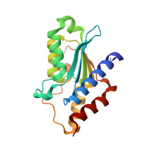
5XKR | pdb_00005xkr

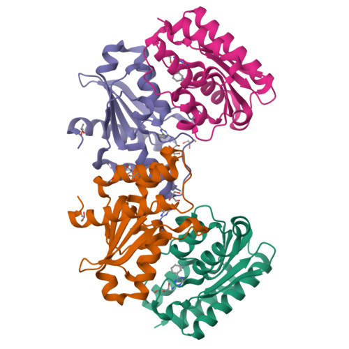
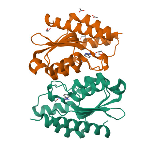
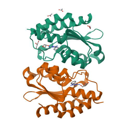
Crystal structure of Msmeg3575 in complex with benzoguanamine


Experimental Data Snapshot

  • Method: X-RAY DIFFRACTION
  • Resolution: 1.38 Å
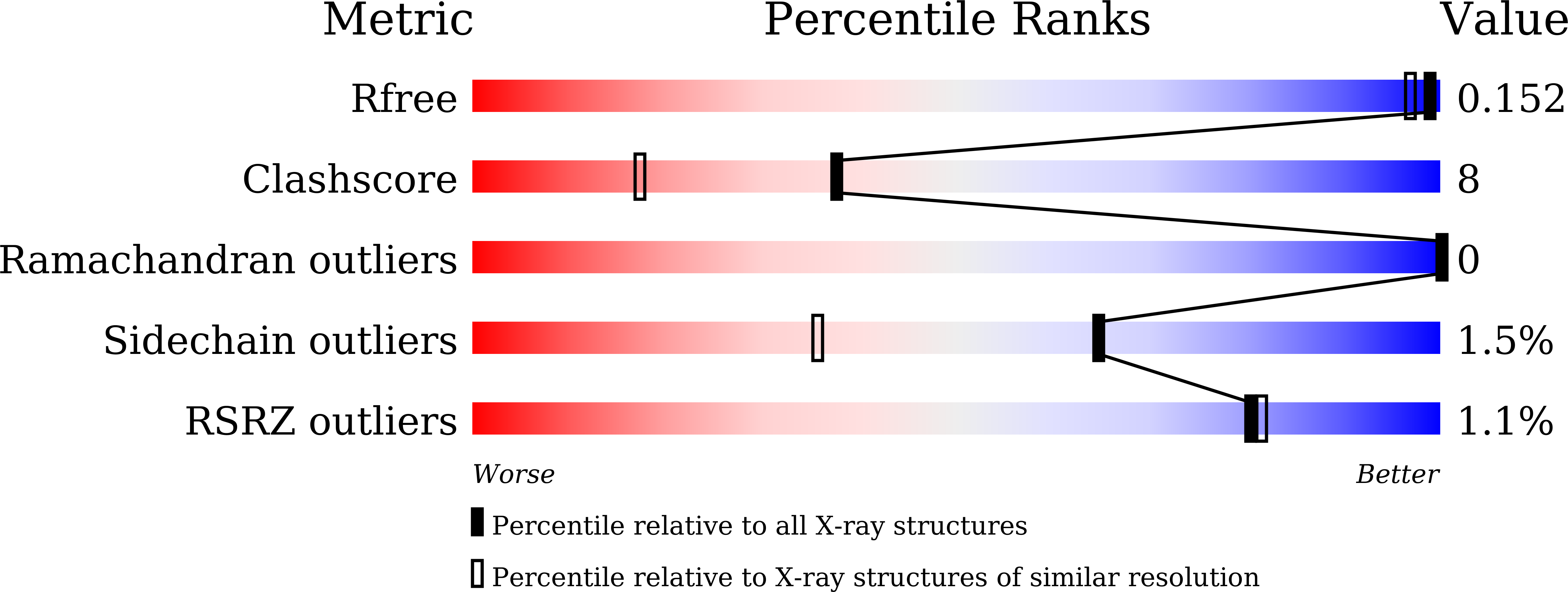
  • R-Value Free: 
    0.153 (Depositor), 0.150 (DCC) 
  • R-Value Work: 
    0.128 (Depositor), 0.130 (DCC) 
  • R-Value Observed: 
    0.129 (Depositor) 

Starting Model: experimental
View more details

wwPDB Validation   3D Report Full Report


Ligand Structure Quality Assessment 

Created with Raphaël 2.3.0Worse 01 BetterLigand structure goodness of fit to experimental dataBest fitted BZEClick on this verticalbar to view details

This is version 1.2 of the entry. See complete history


Literature

Selective Deamination of Mutagens by a Mycobacterial Enzyme

Gaded, V.Anand, R.

(2017) J Am Chem Soc 139: 10762-10768

  • DOI: https://doi.org/10.1021/jacs.7b04967
  • Primary Citation of Related Structures:  
    5XKO, 5XKP, 5XKQ, 5XKR

  • PubMed Abstract: 

    Structure-based methods are powerful tools that are being exploited to unravel new functions with therapeutic advantage. Here, we report the discovery of a new class of deaminases, predominantly found in mycobacterial species that act on the commercially important s-triazine class of compounds. The enzyme Msd from Mycobacterium smegmatis was taken as a representative candidate from an evolutionarily conserved subgroup that possesses high density of Mycobacterium deaminases. Biochemical investigation reveals that Msd specifically acts on mutagenic nucleobases such as 5-azacytosine and isoguanine and does not accept natural bases as substrates. Determination of the X-ray structure of Msd to a resolution of 1.9 Å shows that Msd has fine-tuned its active site such that it is a hybrid of a cytosine as well as a guanine deaminase, thereby conferring Msd the ability to expand its repertoire to both purine and pyrimidine-like mutagens. Mapping of active site residues along with X-ray structures with a series of triazine analogues aids in deciphering the mechanism by which Msd proofreads the base milieu for mutagens. The genome location of the enzyme reveals that Msd is part of a conserved cluster that confers the organism with innate resistance toward select xenobiotics by triggering their efflux.


  • Organizational Affiliation

    Department of Chemistry, Indian Institute of Technology Bombay , Powai, Mumbai 400076, India.


Macromolecules
Find similar proteins by:  (by identity cutoff)  |  3D Structure
Entity ID: 1
MoleculeChains Sequence LengthOrganismDetailsImage
CMP/dCMP deaminase, zinc-binding protein
A, B, C, D
159Mycolicibacterium smegmatis MC2 155Mutation(s): 0 
Gene Names: MSMEG_3575MSMEI_3493
UniProt
Find proteins for A0QY90 (Mycolicibacterium smegmatis (strain ATCC 700084 / mc(2)155))
Explore A0QY90 
Go to UniProtKB:  A0QY90
Entity Groups  
Sequence Clusters30% Identity50% Identity70% Identity90% Identity95% Identity100% Identity
UniProt GroupA0QY90
Sequence Annotations
Expand
  • Reference Sequence
Small Molecules
Ligands 4 Unique
IDChains Name / Formula / InChI Key2D Diagram3D Interactions
BZE
Query on BZE

Download Ideal Coordinates CCD File 
E [auth A],
H [auth B],
P [auth C],
W [auth D]
6-phenyl-1,3,5-triazine-2,4-diamine
C9 H9 N5
GZVHEAJQGPRDLQ-UHFFFAOYSA-N
GOL
Query on GOL

Download Ideal Coordinates CCD File 
G [auth A]
L [auth B]
M [auth B]
N [auth B]
S [auth C]
GLYCEROL
C3 H8 O3
PEDCQBHIVMGVHV-UHFFFAOYSA-N
ZN
Query on ZN

Download Ideal Coordinates CCD File 
F [auth A],
I [auth B],
Q [auth C],
V [auth D]
ZINC ION
Zn
PTFCDOFLOPIGGS-UHFFFAOYSA-N
ACT
Query on ACT

Download Ideal Coordinates CCD File 
J [auth B],
K [auth B],
O [auth C],
R [auth C]
ACETATE ION
C2 H3 O2
QTBSBXVTEAMEQO-UHFFFAOYSA-M
Experimental Data & Validation

Experimental Data

  • Method: X-RAY DIFFRACTION
  • Resolution: 1.38 Å
  • R-Value Free:  0.153 (Depositor), 0.150 (DCC) 
  • R-Value Work:  0.128 (Depositor), 0.130 (DCC) 
  • R-Value Observed: 0.129 (Depositor) 
Space Group: P 1 21 1
Unit Cell:
Length ( Å )Angle ( ˚ )
a = 53.076α = 90
b = 63.446β = 89.99
c = 108.573γ = 90
Software Package:
Software NamePurpose
REFMACrefinement
HKL-3000data reduction
HKL-3000data scaling
Auto-Rickshawphasing

Structure Validation

View Full Validation Report


Ligand Structure Quality Assessment 

Created with Raphaël 2.3.0Worse 01 BetterLigand structure goodness of fit to experimental dataBest fitted BZEClick on this verticalbar to view details

Entry History & Funding Information

Deposition Data


Funding OrganizationLocationGrant Number
Department of Science and Technology, Government of IndiaIndiaEMR/2015/002121 and DST/TM/WTI/2K16/252

Revision History  (Full details and data files)

  • Version 1.0: 2017-08-09
    Type: Initial release
  • Version 1.1: 2017-08-16
    Changes: Database references
  • Version 1.2: 2023-11-22
    Changes: Data collection, Database references, Derived calculations, Refinement description