5XCY

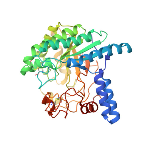
Structure of the cellobiohydrolase Cel6A from Phanerochaete chrysosporium at 1.2 angstrom


Experimental Data Snapshot

  • Method: X-RAY DIFFRACTION
  • Resolution: 1.20 Å
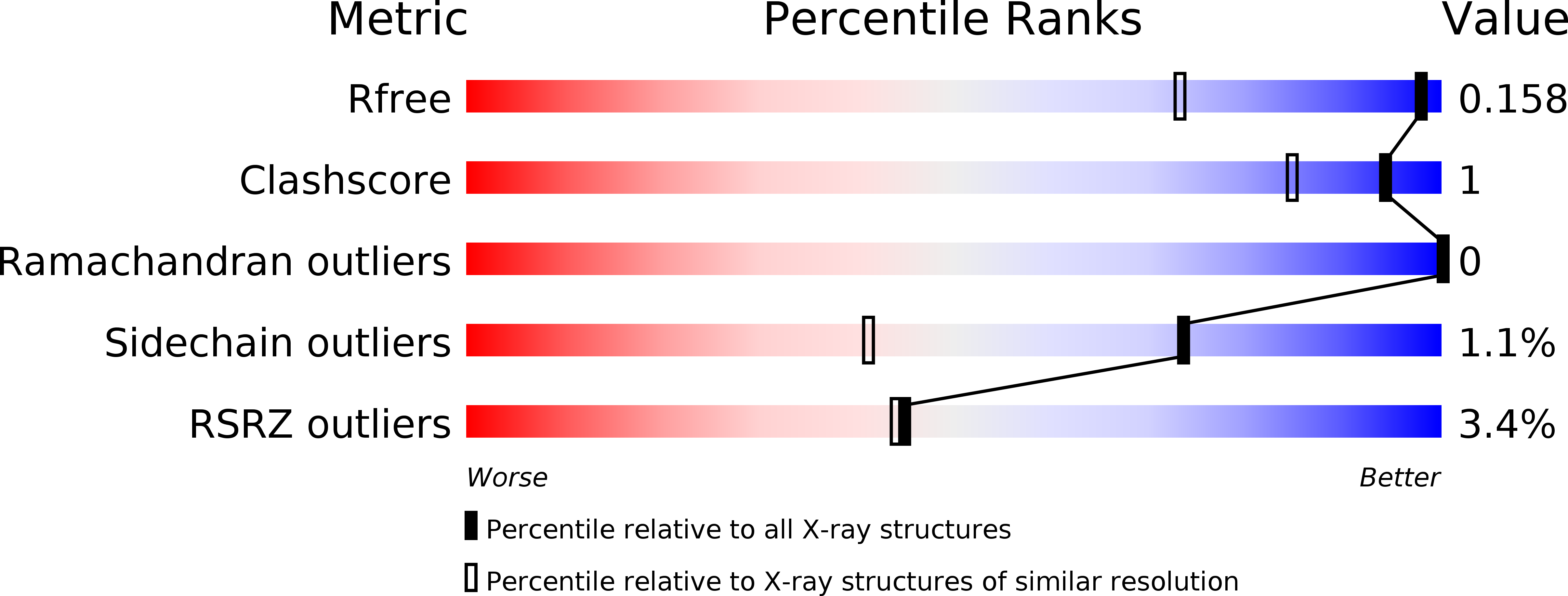
  • R-Value Free: 0.158 
  • R-Value Work: 0.138 
  • R-Value Observed: 0.139 

wwPDB Validation   3D Report Full Report


This is version 1.1 of the entry. See complete history


Literature

Crystal structure of a family 6 cellobiohydrolase from the basidiomycete Phanerochaete chrysosporium

Tachioka, M.Nakamura, A.Ishida, T.Igarashi, K.Samejima, M.

(2017) Acta Crystallogr F Struct Biol Commun 73: 398-403

  • DOI: https://doi.org/10.1107/S2053230X17008093
  • Primary Citation of Related Structures:  
    5XCY, 5XCZ

  • PubMed Abstract: 

    Cellobiohydrolases belonging to glycoside hydrolase family 6 (CBH II, Cel6A) play key roles in the hydrolysis of crystalline cellulose. CBH II from the white-rot fungus Phanerochaete chrysosporium (PcCel6A) consists of a catalytic domain (CD) and a carbohydrate-binding module connected by a linker peptide, like other known fungal cellobiohydrolases. In the present study, the CD of PcCel6A was crystallized without ligands, and p-nitrophenyl β-D-cellotrioside (pNPG3) was soaked into the crystals. The determined structures of the ligand-free and pNPG3-soaked crystals revealed that binding of cellobiose at substrate subsites +1 and +2 induces a conformational change of the N-terminal and C-terminal loops, switching the tunnel-shaped active site from the open to the closed form.


  • Organizational Affiliation

    Department of Biomaterial Sciences, Graduate School of Agricultural and Life Sciences, The University of Tokyo, 1-1-1 Yayoi, Bunkyo-ku, Tokyo 113-8657, Japan.


Macromolecules
Find similar proteins by:  (by identity cutoff)  |  3D Structure
Entity ID: 1
MoleculeChains Sequence LengthOrganismDetailsImage
Glucanase358Phanerodontia chrysosporiumMutation(s): 0 
Gene Names: cel6A
EC: 3.2.1
UniProt
Find proteins for Q02321 (Phanerodontia chrysosporium)
Explore Q02321 
Go to UniProtKB:  Q02321
Entity Groups  
Sequence Clusters30% Identity50% Identity70% Identity90% Identity95% Identity100% Identity
UniProt GroupQ02321
Sequence Annotations
Expand
  • Reference Sequence
Experimental Data & Validation

Experimental Data

  • Method: X-RAY DIFFRACTION
  • Resolution: 1.20 Å
  • R-Value Free: 0.158 
  • R-Value Work: 0.138 
  • R-Value Observed: 0.139 
  • Space Group: P 21 21 21
Unit Cell:
Length ( Å )Angle ( ˚ )
a = 54.712α = 90
b = 67.18β = 90
c = 89.113γ = 90
Software Package:
Software NamePurpose
PHENIXrefinement
HKL-2000data reduction
HKL-2000data scaling
MOLREPphasing

Structure Validation

View Full Validation Report


Entry History & Funding Information

Deposition Data


Funding OrganizationLocationGrant Number
Japanese Ministry of Education, Culture, Sports, and TechnologyJapan24114001
Japanese Ministry of Education, Culture, Sports, and TechnologyJapan24114008
Japan Society for the Promotion of ScienceJapan24380089
Japan Society for the Promotion of ScienceJapan15J10657

Revision History  (Full details and data files)

  • Version 1.0: 2017-07-26
    Type: Initial release
  • Version 1.1: 2024-11-13
    Changes: Data collection, Database references, Structure summary