5XC9 | pdb_00005xc9

Crystal structure of GH45 endoglucanase EG27II at pH8.0, in complex with cellobiose


Experimental Data Snapshot

  • Method: X-RAY DIFFRACTION
  • Resolution: 1.28 Å
  • R-Value Free: 
    0.132 (Depositor), 0.133 (DCC) 
  • R-Value Work: 
    0.105 (Depositor), 0.105 (DCC) 
  • R-Value Observed: 
    0.107 (Depositor) 

Starting Model: experimental
View more details

wwPDB Validation 3D Report Full Report

Validation slider image for 5XC9

This is version 2.2 of the entry. See complete history

Literature

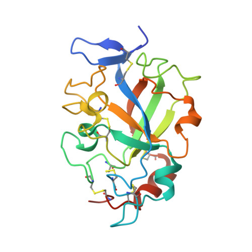
High-resolution crystal structures of the glycoside hydrolase family 45 endoglucanase EG27II from the snail Ampullaria crossean.

Nomura, T.Iwase, H.Saka, N.Takahashi, N.Mikami, B.Mizutani, K.

(2019) Acta Crystallogr D Struct Biol 75: 426-436

  • DOI: https://doi.org/10.1107/S2059798319003000
  • Primary Citation Related Structures: 
    5XBU, 5XBX, 5XC4, 5XC8, 5XC9, 5XCA

  • PubMed Abstract: 

    Although endogenous animal cellulases have great potential for industrial applications such as bioethanol production, few investigations have focused on these enzymes. In this study, the glycoside hydrolase family 45 (GH45) subfamily B endoglucanase EG27II from the snail Ampullaria crossean was expressed using a Pichia pastoris expression system and the crystal structure of the apo form was determined at 1.00 Å resolution; this is the highest resolution structure of an animal endoglucanase. The results showed that EG27II has a double-ψ six-stranded β-barrel and that the structure of EG27II more closely resembles those of subfamily C enzymes than those of subfamily A enzymes. The structure of EG27II complexed with cellobiose was also determined under cryoconditions and at room temperature at three pH values, pH 4.0, 5.5 and 8.0, and no structural changes were found to be associated with the change in pH. The structural comparison and catalytic activity measurements showed that Asp137 and Asn112 function as the catalytic acid and base, respectively, and that Asp27 is also an important residue for catalysis. These high-resolution structures of EG27II provide a large amount of information for structure-based enzyme modification and cell-surface engineering, which will advance biofuel production using animal-derived cellulases.


  • Organizational Affiliation
    • Laboratory of Applied Structural Biology, Division of Applied Life Sciences, Graduate School of Agriculture, Kyoto University, Uji, Kyoto 611-0011, Japan.

Macromolecule Content 

  • Total Structure Weight: 21.6 kDa 
  • Atom Count: 1,708 
  • Modeled Residue Count: 179 
  • Deposited Residue Count: 190 
  • Unique protein chains: 1

Macromolecules

Find similar proteins by:|  3D Structure
Entity ID: 1
MoleculeChains  Sequence LengthOrganismDetailsImage
Endo-beta-1,4-glucanase190Pomacea maculataMutation(s): 0 
Gene Names: EG27II
EC: 3.2.1.4
UniProt
Find proteins for A7KMF0 (Pomacea maculata)
Explore A7KMF0 
Go to UniProtKB:  A7KMF0
Entity Groups
Sequence Clusters30% Identity50% Identity70% Identity90% Identity95% Identity100% Identity
UniProt GroupA7KMF0
Sequence Annotations
Expand
Reference Sequence

Oligosaccharides

Help  
Entity ID: 2
MoleculeChains Length2D Diagram GlycosylationD Interactions
beta-D-glucopyranose-(1-4)-beta-D-glucopyranose
B, C
2N/A
Glycosylation Resources
GlyTouCan: G84824ZO
GlyCosmos: G84824ZO

Biologically Interesting Molecules (External Reference) 

1 Unique

Experimental Data & Validation

Experimental Data

  • Method: X-RAY DIFFRACTION
  • Resolution: 1.28 Å
  • R-Value Free:  0.132 (Depositor), 0.133 (DCC) 
  • R-Value Work:  0.105 (Depositor), 0.105 (DCC) 
  • R-Value Observed: 0.107 (Depositor) 
Space Group: P 21 21 21
Unit Cell:
Length ( Å )Angle ( ˚ )
a = 35.021α = 90
b = 47.278β = 90
c = 92.695γ = 90
Software Package:
Software NamePurpose
PHENIXrefinement
HKL-2000data reduction
HKL-2000data scaling
MOLREPphasing

Structure Validation

View Full Validation Report


Entry History 

& Funding Information

Deposition Data


Funding OrganizationLocationGrant Number
Japan Society for the Promotion of ScienceJapan25450126

Revision History  (Full details and data files)

  • Version 1.0: 2018-03-28
    Type: Initial release
  • Version 1.1: 2019-04-24
    Changes: Data collection, Database references
  • Version 2.0: 2020-07-29
    Type: Remediation
    Reason: Carbohydrate remediation
    Changes: Atomic model, Data collection, Derived calculations, Non-polymer description, Structure summary
  • Version 2.1: 2023-11-22
    Changes: Data collection, Database references, Refinement description, Structure summary
  • Version 2.2: 2024-10-09
    Changes: Structure summary