5X8B | pdb_00005x8b

Crystal structure of ATP-TMP and ADP bound thymidylate kinase from Thermus thermophilus HB8


Experimental Data Snapshot

  • Method: X-RAY DIFFRACTION
  • Resolution: 1.39 Å
  • R-Value Free: 
    0.181 (Depositor), 0.181 (DCC) 
  • R-Value Work: 
    0.144 (Depositor), 0.144 (DCC) 
  • R-Value Observed: 
    0.146 (Depositor) 

Starting Model: experimental
View more details

wwPDB Validation 3D Report Full Report

Validation slider image for 5X8B

Ligand Structure Quality Assessment 


This is version 1.3 of the entry. See complete history

Literature

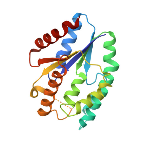
Structural and functional roles of dynamically correlated residues in thymidylate kinase.

Chaudhary, S.K.Jeyakanthan, J.Sekar, K.

(2018) Acta Crystallogr D Struct Biol 74: 341-354

  • DOI: https://doi.org/10.1107/S2059798318002267
  • Primary Citation Related Structures: 
    5X86, 5X8A, 5X8B, 5X8C, 5X8D, 5X8J, 5X8K, 5X8V, 5X98, 5X99, 5XAK, 5XAL, 5XT8

  • PubMed Abstract: 

    Thymidylate kinase is an important enzyme in DNA synthesis. It catalyzes the conversion of thymidine monophosphate to thymidine diphosphate, with ATP as the preferred phosphoryl donor, in the presence of Mg 2+ . In this study, the dynamics of the active site and the communication paths between the substrates, ATP and TMP, are reported for thymidylate kinase from Thermus thermophilus. Conformational changes upon ligand binding and the path for communication between the substrates and the protein are important in understanding the catalytic mechanism of the enzyme. High-resolution X-ray crystal structures of thymidylate kinase in apo and ligand-bound states were solved. This is the first report of structures of binary and ternary complexes of thymidylate kinase with its natural substrates ATP and ATP-TMP, respectively. Distinct conformations of the active-site residues, the P-loop and the LID region observed in the apo and ligand-bound structures revealed that their concerted motion is required for the binding and proper positioning of the substrate TMP. Structural analyses provide an insight into the mode of substrate binding at the active site. The residues involved in communication between the substrates were identified through network analysis using molecular-dynamics simulations. The residues identified showed high sequence conservation across species. Biochemical analyses show that mutations of these residues either resulted in a loss of activity or affected the thermal stability of the protein. Further, molecular-dynamics analyses of mutants suggest that the proper positioning of TMP is important for catalysis. These data also provide an insight into the phosphoryl-transfer mechanism.


  • Organizational Affiliation
    • Department of Physics, Indian Institute of Science, Bangalore 560 012, India.

Macromolecule Content 

  • Total Structure Weight: 45.45 kDa 
  • Atom Count: 3,312 
  • Modeled Residue Count: 376 
  • Deposited Residue Count: 396 
  • Unique protein chains: 1

Macromolecules

Find similar proteins by:|  3D Structure
Entity ID: 1
MoleculeChains  Sequence LengthOrganismDetailsImage
Thymidylate kinase
A, B
198Thermus thermophilus HB8Mutation(s): 0 
Gene Names: tmkTTHA1607
EC: 2.7.4.9
UniProt
Find proteins for Q5SHX3 (Thermus thermophilus (strain ATCC 27634 / DSM 579 / HB8))
Explore Q5SHX3 
Go to UniProtKB:  Q5SHX3
Entity Groups
Sequence Clusters30% Identity50% Identity70% Identity90% Identity95% Identity100% Identity
UniProt GroupQ5SHX3
Sequence Annotations
Expand
Reference Sequence

Small Molecules

Ligands 4 Unique
IDChains Name / Formula / InChI Key2D Diagram3D Interactions
ATP

Query on ATP


Download:Ideal Coordinates CCD File
C [auth A]ADENOSINE-5'-TRIPHOSPHATE
C10 H16 N5 O13 P3
ZKHQWZAMYRWXGA-KQYNXXCUSA-N
ADP

Query on ADP


Download:Ideal Coordinates CCD File
L [auth B]ADENOSINE-5'-DIPHOSPHATE
C10 H15 N5 O10 P2
XTWYTFMLZFPYCI-KQYNXXCUSA-N
TMP

Query on TMP


Download:Ideal Coordinates CCD File
D [auth A]THYMIDINE-5'-PHOSPHATE
C10 H15 N2 O8 P
GYOZYWVXFNDGLU-XLPZGREQSA-N
MG

Query on MG


Download:Ideal Coordinates CCD File
E [auth A]
F [auth A]
G [auth A]
H [auth A]
I [auth B]
E [auth A],
F [auth A],
G [auth A],
H [auth A],
I [auth B],
J [auth B],
K [auth B]
MAGNESIUM ION
Mg
JLVVSXFLKOJNIY-UHFFFAOYSA-N

Experimental Data & Validation

Experimental Data

  • Method: X-RAY DIFFRACTION
  • Resolution: 1.39 Å
  • R-Value Free:  0.181 (Depositor), 0.181 (DCC) 
  • R-Value Work:  0.144 (Depositor), 0.144 (DCC) 
  • R-Value Observed: 0.146 (Depositor) 
Space Group: P 21 21 21
Unit Cell:
Length ( Å )Angle ( ˚ )
a = 47.058α = 90
b = 47.467β = 90
c = 152.453γ = 90
Software Package:
Software NamePurpose
REFMACrefinement
HKL-2000data reduction
Aimlessdata scaling
PHASERphasing

Structure Validation

View Full Validation Report


Ligand Structure Quality Assessment 


Entry History 

Deposition Data

Revision History  (Full details and data files)

  • Version 1.0: 2018-03-14
    Type: Initial release
  • Version 1.1: 2018-04-18
    Changes: Data collection, Database references
  • Version 1.2: 2018-11-21
    Changes: Data collection, Database references
  • Version 1.3: 2023-11-22
    Changes: Data collection, Database references, Derived calculations, Refinement description