5X3S | pdb_00005x3s

Crystal structure of mouse Plk1-PBD in complex with phosphopeptide from HEF1 (799-809)


Experimental Data Snapshot

  • Method: X-RAY DIFFRACTION
  • Resolution: 2.90 Å
  • R-Value Free: 
    0.244 (Depositor), 0.240 (DCC) 
  • R-Value Work: 
    0.203 (Depositor), 0.202 (DCC) 
  • R-Value Observed: 
    0.204 (Depositor) 

Starting Model: experimental
View more details

wwPDB Validation 3D Report Full Report

Validation slider image for 5X3S

This is version 1.3 of the entry. See complete history

Literature

Phosphorylation of human enhancer filamentation 1 (HEF1) stimulates interaction with Polo-like kinase 1 leading to HEF1 localization to focal adhesions.

Lee, K.H.Hwang, J.A.Kim, S.O.Kim, J.H.Shin, S.C.Kim, E.E.Lee, K.S.Rhee, K.Jeon, B.H.Bang, J.K.Cha-Molstad, H.Soung, N.K.Jang, J.H.Ko, S.K.Lee, H.G.Ahn, J.S.Kwon, Y.T.Kim, B.Y.

(2018) J Biological Chem 293: 847-862

  • DOI: https://doi.org/10.1074/jbc.M117.802587
  • Primary Citation Related Structures: 
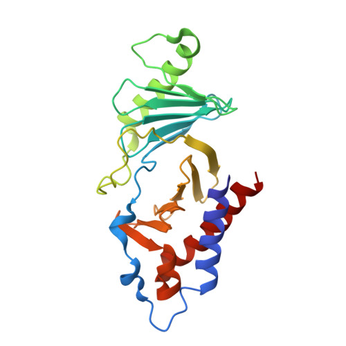
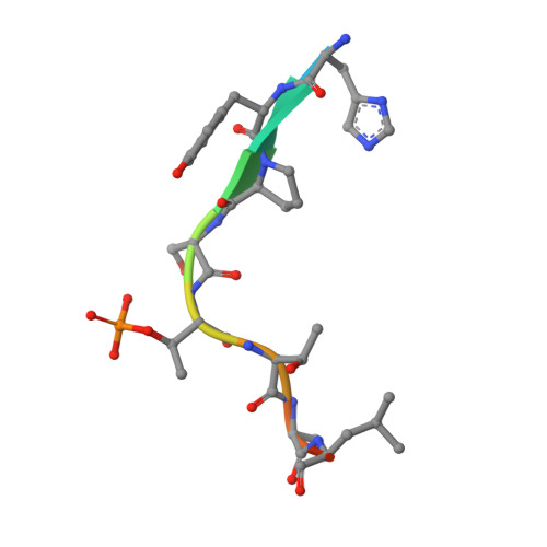
    5X3S

  • PubMed Abstract: 

    Elevated expression of human enhancer filamentation 1 (HEF1; also known as NEDD9 or Cas-L) is an essential stimulus for the metastatic process of various solid tumors. This process requires HEF1 localization to focal adhesions (FAs). Although the association of HEF1 with FAs is considered to play a role in cancer cell migration, the mechanism targeting HEF1 to FAs remains unclear. Moreover, up-regulation of Polo-like kinase 1 (Plk1) positively correlates with human cancer metastasis, yet how Plk1 deregulation promotes metastasis remains elusive. Here, we report that casein kinase 1δ (CK1δ) phosphorylates HEF1 at Ser-780 and Thr-804 and that these phosphorylation events promote a physical interaction between Plk1 and HEF1. We found that this interaction is critical for HEF1 translocation to FAs and for inducing migration of HeLa cells. Plk1-docking phosphoepitopes were mapped/confirmed in HEF1 by various methods, including X-ray crystallography, and mutated for functional analysis in HeLa cells. In summary, our results reveal the role of a phosphorylation-dependent HEF1-Plk1 complex in HEF1 translocation to FAs to induce cell migration. Our findings provide critical mechanistic insights into the HEF1-Plk1 complex-dependent localization of HEF1 to FAs underlying the metastatic process and may therefore contribute to the development of new cancer therapies.


  • Organizational Affiliation
    • From the World Class Institute, Anticancer Agent Research Center, Korea Research Institute of Bioscience and Biotechnology, 30 Yeongudanji-ro, Ochang, Cheongwon, Chungbuk 28116, Korea, leekh@kribb.re.kr.

Macromolecule Content 

  • Total Structure Weight: 54.48 kDa 
  • Atom Count: 3,753 
  • Modeled Residue Count: 462 
  • Deposited Residue Count: 470 
  • Unique protein chains: 2

Macromolecules

Find similar proteins by:|  3D Structure
Entity ID: 1
MoleculeChains  Sequence LengthOrganismDetailsImage
Serine/threonine-protein kinase PLK1A [auth B],
B [auth A]
224Mus musculusMutation(s): 0 
Gene Names: Plk1Plk
EC: 2.7.11.21
UniProt & NIH Common Fund Data Resources
Find proteins for Q07832 (Mus musculus)
Explore Q07832 
Go to UniProtKB:  Q07832
IMPC:  MGI:97621
Entity Groups
Sequence Clusters30% Identity50% Identity70% Identity90% Identity95% Identity100% Identity
UniProt GroupQ07832
Sequence Annotations
Expand
Reference Sequence
Find similar proteins by:  Sequence   |   3D Structure  
Entity ID: 2
MoleculeChains  Sequence LengthOrganismDetailsImage
Peptide from Enhancer of filamentation 1
C, D
11Homo sapiensMutation(s): 0 
UniProt & NIH Common Fund Data Resources
Find proteins for Q14511 (Homo sapiens)
Explore Q14511 
Go to UniProtKB:  Q14511
PHAROS:  Q14511
GTEx:  ENSG00000111859 
Entity Groups
Sequence Clusters30% Identity50% Identity70% Identity90% Identity95% Identity100% Identity
UniProt GroupQ14511
Sequence Annotations
Expand
Reference Sequence

Small Molecules

Modified Residues  1 Unique
IDChains TypeFormula2D DiagramParent
TPO
Query on TPO
C, D
L-PEPTIDE LINKINGC4 H10 N O6 PTHR

Experimental Data & Validation

Experimental Data

  • Method: X-RAY DIFFRACTION
  • Resolution: 2.90 Å
  • R-Value Free:  0.244 (Depositor), 0.240 (DCC) 
  • R-Value Work:  0.203 (Depositor), 0.202 (DCC) 
  • R-Value Observed: 0.204 (Depositor) 
Space Group: P 1 21 1
Unit Cell:
Length ( Å )Angle ( ˚ )
a = 57.564α = 90
b = 59.402β = 99.47
c = 72.748γ = 90
Software Package:
Software NamePurpose
PHENIXrefinement
HKL-2000data scaling
PDB_EXTRACTdata extraction
PHASERphasing
HKLdata scaling

Structure Validation

View Full Validation Report


Entry History 

Deposition Data

Revision History  (Full details and data files)

  • Version 1.0: 2017-12-20
    Type: Initial release
  • Version 1.1: 2018-01-31
    Changes: Database references
  • Version 1.2: 2023-11-22
    Changes: Data collection, Database references, Refinement description
  • Version 1.3: 2024-11-06
    Changes: Structure summary