5X0F | pdb_00005x0f

Free serine kinase (E30A mutant) in complex with AMP


Experimental Data Snapshot

  • Method: X-RAY DIFFRACTION
  • Resolution: 1.76 Å
  • R-Value Free: 
    0.261 (Depositor), 0.271 (DCC) 
  • R-Value Work: 
    0.223 (Depositor) 
  • R-Value Observed: 
    0.225 (Depositor) 

Starting Model: experimental
View more details

wwPDB Validation 3D Report Full Report

Validation slider image for 5X0F

Ligand Structure Quality Assessment 


This is version 1.2 of the entry. See complete history

Literature

Structural Study on the Reaction Mechanism of a Free Serine Kinase Involved in Cysteine Biosynthesis

Nagata, R.Fujihashi, M.Kawamura, H.Sato, T.Fujita, T.Atomi, H.Miki, K.

(2017) ACS Chem Biol 12: 1514-1523

  • DOI: https://doi.org/10.1021/acschembio.7b00064
  • Primary Citation Related Structures: 
    5X0B, 5X0E, 5X0F, 5X0G, 5X0J, 5X0K

  • PubMed Abstract: 

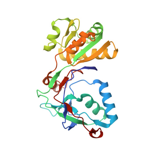
    A free serine kinase (SerK) is involved in l-cysteine biosynthesis in the hyperthermophilic archaeon Thermococcus kodakarensis. The enzyme converts ADP and l-serine (Ser) into AMP and O-phospho-l-serine (Sep), which is a precursor of l-cysteine. SerK is the first identified enzyme that phosphorylates free serine, while serine/threonine protein kinases have been well studied. SerK displays low sequence similarities to known kinases, suggesting that its reaction mechanism is different from those of known kinases. Here, we determined the crystal structures of SerK from T. kodakarensis (Tk-SerK). The overall structure is divided into two domains. A large cleft is found between the two domains in the AMP complex and in the ADP complex. The cleft is closed in the ternary product complex (Sep, AMP, and Tk-SerK) and may also be in the ternary substrate complex (Ser, ADP, and Tk-SerK). The closure may reorient the carboxyl group of E30 near to the Oγ atom of Ser. The Oγ atom is considered to be deprotonated by E30 and to attack the β-phosphate of ADP to form Sep. The substantial decrease in the activity of the E30A mutant is consistent with this mechanism. Our structures also revealed the residues that contribute to the ligand binding. The conservation of these residues in uncharacterized proteins from bacteria may raise the possibility of the presence of free Ser kinases not only in archaea but also in bacteria.


  • Organizational Affiliation
    • Department of Chemistry, Graduate School of Science, Kyoto University , Sakyo-ku, Kyoto 606-8502, Japan.

Macromolecule Content 

  • Total Structure Weight: 28.03 kDa 
  • Atom Count: 2,017 
  • Modeled Residue Count: 241 
  • Deposited Residue Count: 242 
  • Unique protein chains: 1

Macromolecules

Find similar proteins by:|  3D Structure
Entity ID: 1
MoleculeChains  Sequence LengthOrganismDetailsImage
Free serine kinase242Thermococcus kodakarensis KOD1Mutation(s): 1 
Gene Names: TK0378
EC: 2.7.1.226
UniProt
Find proteins for Q5JD03 (Thermococcus kodakarensis (strain ATCC BAA-918 / JCM 12380 / KOD1))
Explore Q5JD03 
Go to UniProtKB:  Q5JD03
Entity Groups
Sequence Clusters30% Identity50% Identity70% Identity90% Identity95% Identity100% Identity
UniProt GroupQ5JD03
Sequence Annotations
Expand
Reference Sequence

Small Molecules

Ligands 1 Unique
IDChains Name / Formula / InChI Key2D Diagram3D Interactions
AMP

Query on AMP


Download:Ideal Coordinates CCD File
B [auth A]ADENOSINE MONOPHOSPHATE
C10 H14 N5 O7 P
UDMBCSSLTHHNCD-KQYNXXCUSA-N

Experimental Data & Validation

Experimental Data

  • Method: X-RAY DIFFRACTION
  • Resolution: 1.76 Å
  • R-Value Free:  0.261 (Depositor), 0.271 (DCC) 
  • R-Value Work:  0.223 (Depositor) 
  • R-Value Observed: 0.225 (Depositor) 
Space Group: P 21 21 2
Unit Cell:
Length ( Å )Angle ( ˚ )
a = 39.903α = 90
b = 70.032β = 90
c = 88.381γ = 90
Software Package:
Software NamePurpose
REFMACrefinement
HKL-2000data scaling
REFMACphasing

Structure Validation

View Full Validation Report


Ligand Structure Quality Assessment 


Entry History 

Deposition Data

Revision History  (Full details and data files)

  • Version 1.0: 2017-04-12
    Type: Initial release
  • Version 1.1: 2017-07-05
    Changes: Database references
  • Version 1.2: 2023-11-22
    Changes: Data collection, Database references, Refinement description