5WZE | pdb_00005wze

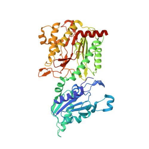
The structure of Pseudomonas aeruginosa aminopeptidase PepP


Experimental Data Snapshot

  • Method: X-RAY DIFFRACTION
  • Resolution: 1.78 Å
  • R-Value Free: 
    0.221 (Depositor), 0.211 (DCC) 
  • R-Value Work: 
    0.210 (Depositor) 
  • R-Value Observed: 
    0.211 (Depositor) 

Starting Model: experimental
View more details

wwPDB Validation 3D Report Full Report

Validation slider image for 5WZE

This is version 1.1 of the entry. See complete history. 

Literature

Structure-Function Relationship of Aminopeptidase P from Pseudomonas aeruginosa.

Peng, C.T., Liu, L., Li, C.C., He, L.H., Li, T., Shen, Y.L., Gao, C., Wang, N.Y., Xia, Y., Zhu, Y.B., Song, Y.J., Lei, Q., Yu, L.T., Bao, R.

(2017) Front Microbiol 8: 2385-2385

  • DOI: https://doi.org/10.3389/fmicb.2017.02385
  • Primary Citation Related Structures: 
    5WZE

  • PubMed Abstract: 

    PepP is a virulence-associated gene in Pseudomonas aeruginosa , making it an attractive target for anti -P. aeruginosa drug development. The encoded protein, aminopeptidases P (Pa-PepP), is a type of X-prolyl peptidase that possesses diverse biological functions. The crystal structure verified its canonical pita-bread fold and functional tetrameric assembly, and the functional studies measured the influences of different metal ions on the activity. A trimetal manganese cluster was observed at the active site, elucidating the mechanism of inhibition by metal ions. Additionally, a loop extending from the active site appeared to be important for specific large-substrate binding. Based on the structural comparison and bacterial invasion assays, we showed that this non-conserved surface loop was critical for P. aeruginosa virulence. Taken together, these findings can extend our understanding of the catalytic mechanism and virulence-related functions of Pa-PepP and provide a solid foundation for the design of specific inhibitors against pathogenic-bacterial infections.


  • Organizational Affiliation: 
    • Pharmaceutical and Biological Engineering Department, School of Chemical Engineering, Sichuan University, Chengdu, China.

Macromolecule Content 

  • Total Structure Weight: 206.09 kDa 
  • Atom Count: 15,378 
  • Modeled Residue Count: 1,777 
  • Deposited Residue Count: 1,808 
  • Unique protein chains: 1

Macromolecules

Find similar proteins by:|  3D Structure
Entity ID: 1
MoleculeChains  Sequence LengthOrganismDetailsImage
Aminopeptidase P
A, B, C, D
452Pseudomonas aeruginosa PAO1Mutation(s): 0 
Gene Names: pepP, PA5224
EC: 3.4.11.9
UniProt
Find proteins for Q9HTW6 (Pseudomonas aeruginosa (strain ATCC 15692 / DSM 22644 / CIP 104116 / JCM 14847 / LMG 12228 / 1C / PRS 101 / PAO1))
Explore Q9HTW6 
Go to UniProtKB:  Q9HTW6
Entity Groups
Sequence Clusters30% Identity50% Identity70% Identity90% Identity95% Identity100% Identity
UniProt GroupQ9HTW6
Sequence Annotations
Expand
Reference Sequence

Small Molecules

Ligands 9 Unique
IDChains Name / Formula / InChI Key2D Diagram3D Interactions
PGE

Query on PGE


Download:Ideal Coordinates CCD File
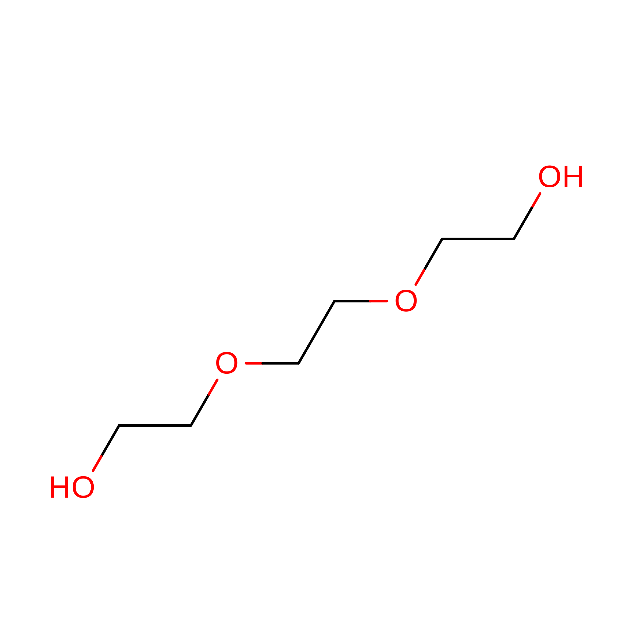
P [auth B]TRIETHYLENE GLYCOL
C6 H14 O4
ZIBGPFATKBEMQZ-UHFFFAOYSA-N
PRO

Query on PRO


Download:Ideal Coordinates CCD File
CA [auth C],
H [auth A],
RA [auth D],
S [auth B]
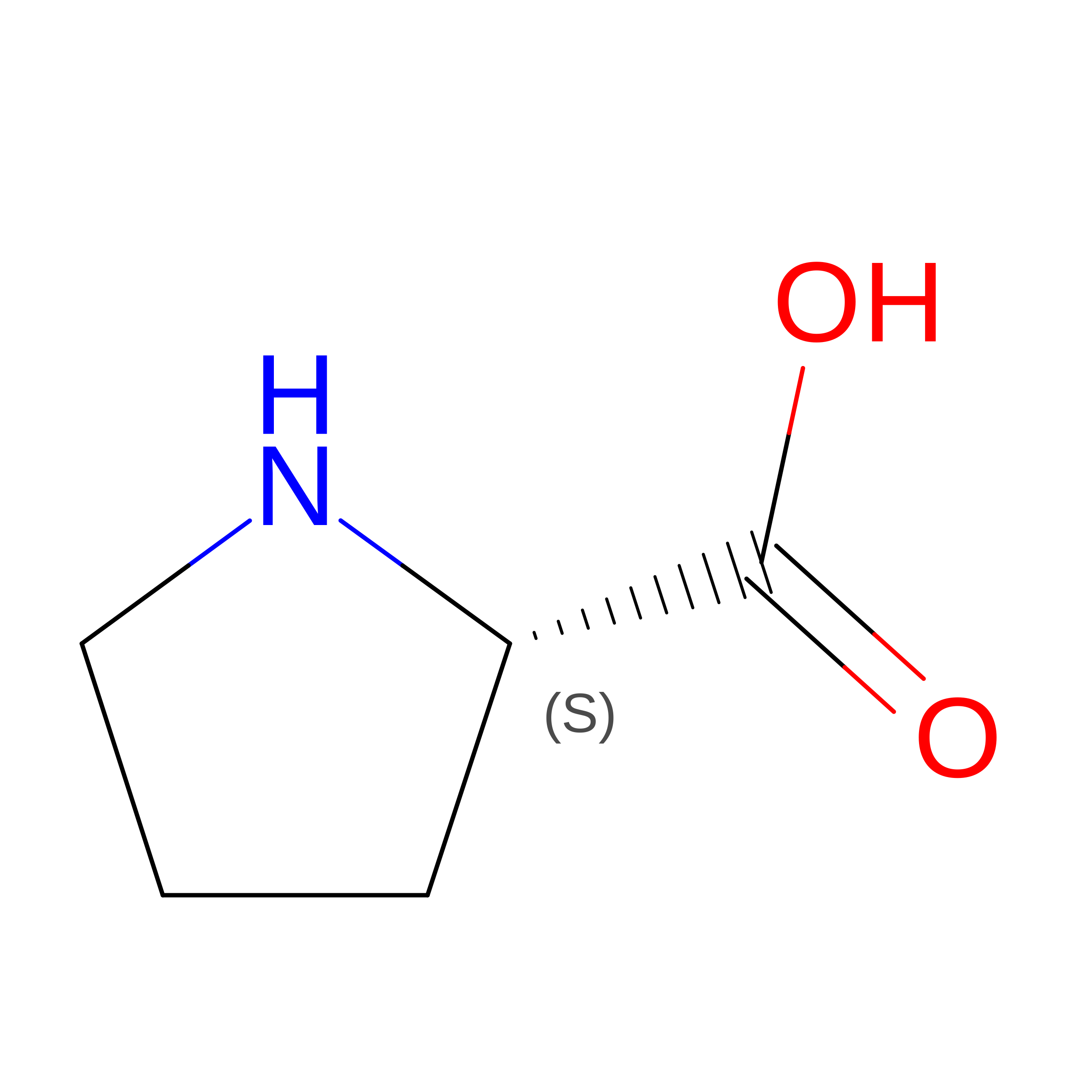
PROLINE
C5 H9 N O2
ONIBWKKTOPOVIA-BYPYZUCNSA-N
SO4

Query on SO4


Download:Ideal Coordinates CCD File
G [auth A],
LA [auth D],
MA [auth D]
SULFATE ION
O4 S
QAOWNCQODCNURD-UHFFFAOYSA-L
GOL

Query on GOL


Download:Ideal Coordinates CCD File
F [auth A],
IA [auth D],
JA [auth D],
KA [auth D]
GLYCEROL
C3 H8 O3
PEDCQBHIVMGVHV-UHFFFAOYSA-N
ALA

Query on ALA


Download:Ideal Coordinates CCD File
I [auth A],
T [auth B]
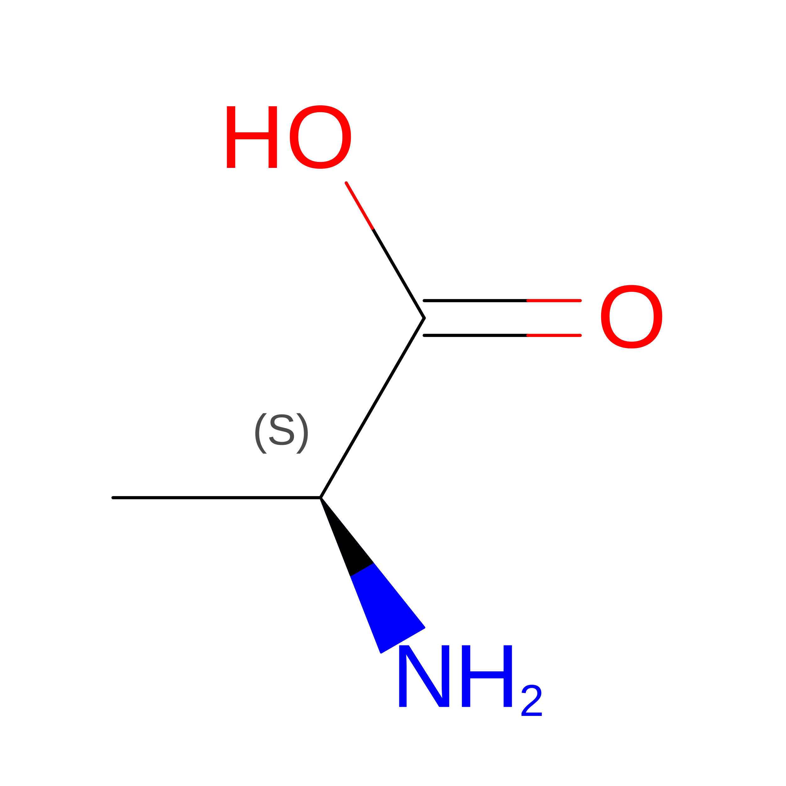
ALANINE
C3 H7 N O2
QNAYBMKLOCPYGJ-REOHCLBHSA-N
EDO

Query on EDO


Download:Ideal Coordinates CCD File
AA [auth C],
BA [auth C],
NA [auth D],
Q [auth B],
R [auth B]
1,2-ETHANEDIOL
C2 H6 O2
LYCAIKOWRPUZTN-UHFFFAOYSA-N
MN

Query on MN


Download:Ideal Coordinates CCD File
FA [auth C]
GA [auth C]
HA [auth C]
K [auth A]
L [auth A]
FA [auth C],
GA [auth C],
HA [auth C],
K [auth A],
L [auth A],
M [auth A],
OA [auth D],
PA [auth D],
QA [auth D],
W [auth B],
X [auth B],
Y [auth B]
MANGANESE (II) ION
Mn
WAEMQWOKJMHJLA-UHFFFAOYSA-N
CA

Query on CA


Download:Ideal Coordinates CCD File
DA [auth C],
EA [auth C],
J [auth A],
U [auth B],
V [auth B]
CALCIUM ION
Ca
BHPQYMZQTOCNFJ-UHFFFAOYSA-N
NA

Query on NA


Download:Ideal Coordinates CCD File
E [auth A],
N [auth B],
O [auth B],
Z [auth C]
SODIUM ION
Na
FKNQFGJONOIPTF-UHFFFAOYSA-N

Experimental Data & Validation

Experimental Data

  • Method: X-RAY DIFFRACTION
  • Resolution: 1.78 Å
  • R-Value Free:  0.221 (Depositor), 0.211 (DCC) 
  • R-Value Work:  0.210 (Depositor) 
  • R-Value Observed: 0.211 (Depositor) 
Space Group: P 21 21 21
Unit Cell:
Length ( Å )Angle ( ˚ )
a = 111.197α = 90
b = 123.432β = 90
c = 149.485γ = 90
Software Package:
Software NamePurpose
PHENIXrefinement
HKL-2000data reduction
HKL-2000data scaling
PHASERphasing

Structure Validation

View Full Validation Report


Entry History 

Deposition Data

Revision History  (Full details and data files)

  • Version 1.0: 2018-01-17
    Type: Initial release
  • Version 1.1: 2023-11-22
    Changes: Data collection, Database references, Derived calculations, Refinement description