5WYA | pdb_00005wya

Structure of amino acid racemase, 2.65 A


Experimental Data Snapshot

  • Method: X-RAY DIFFRACTION
  • Resolution: 2.65 Å
  • R-Value Free: 
    0.222 (Depositor), 0.220 (DCC) 
  • R-Value Work: 
    0.175 (Depositor), 0.182 (DCC) 
  • R-Value Observed: 
    0.178 (Depositor) 

wwPDB Validation 3D Report Full Report

Validation slider image for 5WYA

Ligand Structure Quality Assessment 


This is version 1.2 of the entry. See complete history

Literature

Crystal structure of the novel amino-acid racemase isoleucine 2-epimerase from Lactobacillus buchneri.

Hayashi, J.Mutaguchi, Y.Minemura, Y.Nakagawa, N.Yoneda, K.Ohmori, T.Ohshima, T.Sakuraba, H.

(2017) Acta Crystallogr D Struct Biol 73: 428-437

  • DOI: https://doi.org/10.1107/S2059798317005332
  • Primary Citation Related Structures: 
    4YSN, 4YSV, 5WYA, 5WYF

  • PubMed Abstract: 

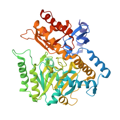
    Crystal structures of Lactobacillus buchneri isoleucine 2-epimerase, a novel branched-chain amino-acid racemase, were determined for the enzyme in the apo form, in complex with pyridoxal 5'-phosphate (PLP), in complex with N-(5'-phosphopyridoxyl)-L-isoleucine (PLP-L-Ile) and in complex with N-(5'-phosphopyridoxyl)-D-allo-isoleucine (PLP-D-allo-Ile) at resolutions of 2.77, 1.94, 2.65 and 2.12 Å, respectively. The enzyme assembled as a tetramer, with each subunit being composed of N-terminal, C-terminal and large PLP-binding domains. The active-site cavity in the apo structure was much more solvent-accessible than that in the PLP-bound structure. This indicates that a marked structural change occurs around the active site upon binding of PLP that provides a solvent-inaccessible environment for the enzymatic reaction. The main-chain coordinates of the L. buchneri isoleucine 2-epimerase monomer showed a notable similarity to those of α-amino-ℇ-caprolactam racemase from Achromobactor obae and γ-aminobutyrate aminotransferase from Escherichia coli. However, the amino-acid residues involved in substrate binding in those two enzymes are only partially conserved in L. buchneri isoleucine 2-epimerase, which may account for the differences in substrate recognition by the three enzymes. The structures bound with reaction-intermediate analogues (PLP-L-Ile and PLP-D-allo-Ile) and site-directed mutagenesis suggest that L-isoleucine epimerization proceeds through abstraction of the α-hydrogen of the substrate by Lys280, while Asp222 serves as the catalytic residue adding an α-hydrogen to the quinonoid intermediate to form D-allo-isoleucine.


  • Organizational Affiliation
    • Department of Applied Biological Science, Faculty of Agriculture, Kagawa University, 2393 Ikenobe, Miki-cho, Kita-gun, Kagawa 761-0795, Japan.

Macromolecule Content 

  • Total Structure Weight: 200.24 kDa 
  • Atom Count: 14,376 
  • Modeled Residue Count: 1,758 
  • Deposited Residue Count: 1,800 
  • Unique protein chains: 1

Macromolecules

Find similar proteins by:|  3D Structure
Entity ID: 1
MoleculeChains  Sequence LengthOrganismDetailsImage
Isoleucine 2-epimerase
A, B, C, D
450Lentilactobacillus buchneriMutation(s): 0 
EC: 5.1.1.21
UniProt
Find proteins for M1GRN3 (Lentilactobacillus buchneri)
Explore M1GRN3 
Go to UniProtKB:  M1GRN3
Entity Groups
Sequence Clusters30% Identity50% Identity70% Identity90% Identity95% Identity100% Identity
UniProt GroupM1GRN3
Sequence Annotations
Expand
Reference Sequence

Small Molecules

Ligands 2 Unique
IDChains Name / Formula / InChI Key2D Diagram3D Interactions
7VO

Query on 7VO


Download:Ideal Coordinates CCD File
E [auth A],
J [auth B],
O [auth C],
R [auth D]
(2S,3S)-3-methyl-2-[[2-methyl-3-oxidanyl-5-(phosphonooxymethyl)pyridin-4-yl]methylamino]pentanoic acid
C14 H23 N2 O7 P
GZZDWFDWHXPWJK-UFBFGSQYSA-N
DMS

Query on DMS


Download:Ideal Coordinates CCD File
F [auth A]
G [auth A]
H [auth A]
I [auth A]
K [auth B]
F [auth A],
G [auth A],
H [auth A],
I [auth A],
K [auth B],
L [auth B],
M [auth B],
N [auth C],
P [auth C],
Q [auth C],
S [auth D]
DIMETHYL SULFOXIDE
C2 H6 O S
IAZDPXIOMUYVGZ-UHFFFAOYSA-N

Experimental Data & Validation

Experimental Data

  • Method: X-RAY DIFFRACTION
  • Resolution: 2.65 Å
  • R-Value Free:  0.222 (Depositor), 0.220 (DCC) 
  • R-Value Work:  0.175 (Depositor), 0.182 (DCC) 
  • R-Value Observed: 0.178 (Depositor) 
Space Group: P 21 21 21
Unit Cell:
Length ( Å )Angle ( ˚ )
a = 61.091α = 90
b = 160.681β = 90
c = 186.28γ = 90
Software Package:
Software NamePurpose
HKL-2000data collection
HKL-2000data scaling
MOLREPphasing
REFMACrefinement
PDB_EXTRACTdata extraction
HKL-2000data reduction
HKLdata reduction
HKLdata scaling

Structure Validation

View Full Validation Report


Ligand Structure Quality Assessment 


Entry History 

Deposition Data

Revision History  (Full details and data files)

  • Version 1.0: 2017-04-19
    Type: Initial release
  • Version 1.1: 2017-05-17
    Changes: Database references
  • Version 1.2: 2024-03-20
    Changes: Data collection, Database references