5WXO

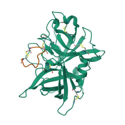
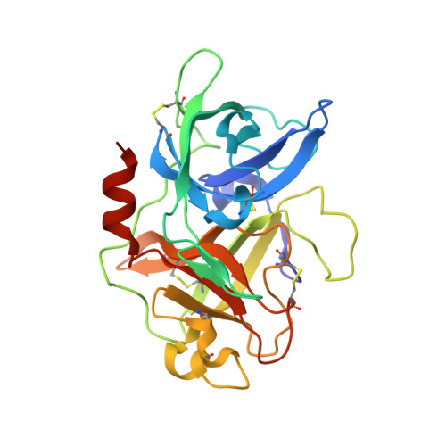
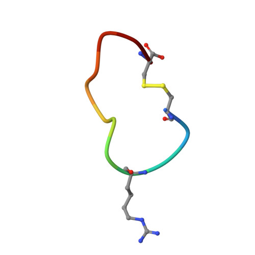
Crystal structure of uPA in complex with upain-2-2-W3A


Experimental Data Snapshot

  • Method: X-RAY DIFFRACTION
  • Resolution: 1.64 Å
  • R-Value Free: 0.221 
  • R-Value Work: 0.180 
  • R-Value Observed: 0.182 

Starting Model: experimental
View more details

wwPDB Validation   3D Report Full Report


This is version 1.2 of the entry. See complete history


Literature

Cleavage of peptidic inhibitors by target protease is caused by peptide conformational transition.

Jiang, L.Oldenburg, E.Kromann-Hansen, T.Xu, P.Jensen, J.K.Andreasen, P.A.Huang, M.

(2018) Biochim Biophys Acta 1862: 2017-2023

  • DOI: https://doi.org/10.1016/j.bbagen.2018.06.016
  • Primary Citation of Related Structures:  
    5WXF, 5WXO, 5WXP

  • PubMed Abstract: 

    Some peptide sequences can behave as either substrates or inhibitors of serine proteases. Working with a cyclic peptidic inhibitor of the serine protease urokinase-type plasminogen activator (uPA), we have now demonstrated a new mechanism for an inhibitor-to-substrate switch. The peptide, CSWRGLENHAAC (upain-2), is a competitive inhibitor of human uPA, but is also slowly converted to a substrate in which the bond between Arg 4 and Gly 5 (the P1-P1' bond) is cleaved. Substituting the P2 residue Trp 3 to an Ala or substituting the P1 Arg 4 residue with 4-guanidino-phenylalanine strongly increased the substrate cleavage rate. We studied the structural basis for the inhibitor-to-substrate switch by determining the crystal structures of the various peptide variants in complex with the catalytic domain of uPA. While the slowly cleaved peptides bound clearly in inhibitory mode, with the oxyanion hole blocked by the side chain of the P3' residue Glu 7 , peptides behaving essentially as substrates with a much accelerated rate of cleavage was observed to be bound to the enzyme in substrate mode. Our analysis reveals that the inhibitor-to-substrate switch was associated with a 7 Å translocation of the P2 residue, and we conclude that the inhibitor-to-substrate switch of upain-2 is a result of a major conformational change in the enzyme-bound state of the peptide. This conclusion is in contrast to findings with so-called standard mechanism inhibitors in which the inhibitor-to-substrate switch is linked to minor conformational changes in the backbone of the inhibitory peptide stretch.


  • Organizational Affiliation

    College of Chemistry, Fuzhou University, 350116, China.


Macromolecules
Find similar proteins by:  (by identity cutoff)  |  3D Structure
Entity ID: 1
MoleculeChains Sequence LengthOrganismDetailsImage
Urokinase-type plasminogen activator chain BA [auth U]253Homo sapiensMutation(s): 2 
Gene Names: PLAU
EC: 3.4.21.73
UniProt & NIH Common Fund Data Resources
Find proteins for P00749 (Homo sapiens)
Explore P00749 
Go to UniProtKB:  P00749
PHAROS:  P00749
GTEx:  ENSG00000122861 
Entity Groups  
Sequence Clusters30% Identity50% Identity70% Identity90% Identity95% Identity100% Identity
UniProt GroupP00749
Sequence Annotations
Expand
  • Reference Sequence

Find similar proteins by:  Sequence   |   3D Structure  

Entity ID: 2
MoleculeChains Sequence LengthOrganismDetailsImage
upain-2-2-W3A peptideB [auth P]12Phage display vector pTDispMutation(s): 0 
Entity Groups  
Sequence Clusters30% Identity50% Identity70% Identity90% Identity95% Identity100% Identity
Sequence Annotations
Expand
  • Reference Sequence
Small Molecules
Modified Residues  1 Unique
IDChains TypeFormula2D DiagramParent
HRG
Query on HRG
B [auth P]L-PEPTIDE LINKINGC7 H16 N4 O2ARG
Experimental Data & Validation

Experimental Data

  • Method: X-RAY DIFFRACTION
  • Resolution: 1.64 Å
  • R-Value Free: 0.221 
  • R-Value Work: 0.180 
  • R-Value Observed: 0.182 
  • Space Group: H 3
Unit Cell:
Length ( Å )Angle ( ˚ )
a = 121.07α = 90
b = 121.07β = 90
c = 42.88γ = 120
Software Package:
Software NamePurpose
REFMACrefinement
HKL-2000data reduction
HKL-2000data scaling
MOLREPphasing

Structure Validation

View Full Validation Report


Entry History 

Deposition Data

Revision History  (Full details and data files)

  • Version 1.0: 2018-07-11
    Type: Initial release
  • Version 1.1: 2018-07-18
    Changes: Data collection, Database references
  • Version 1.2: 2023-11-22
    Changes: Data collection, Database references, Derived calculations, Refinement description