5WWN | pdb_00005wwn

Crystal structure of Tsr1


Experimental Data Snapshot

  • Method: X-RAY DIFFRACTION
  • Resolution: 2.81 Å
  • R-Value Free: 
    0.274 (Depositor), 0.272 (DCC) 
  • R-Value Work: 
    0.214 (Depositor), 0.218 (DCC) 
  • R-Value Observed: 
    0.217 (Depositor) 

wwPDB Validation 3D Report Full Report

Validation slider image for 5WWN

This is version 1.1 of the entry. See complete history

Literature

Molecular architecture of the 90S small subunit pre-ribosome

Sun, Q.Zhu, X.Qi, J.An, W.Lan, P.Tan, D.Chen, R.Wang, B.Zheng, S.Zhang, C.Chen, X.Zhang, W.Chen, J.Dong, M.Q.Ye, K.

(2017) Elife 6

  • DOI: https://doi.org/10.7554/eLife.22086
  • Primary Citation Related Structures: 
    5WWN, 5WWO, 5WXL, 5WXM, 5WY3, 5WYJ, 5WYK, 5WYL

  • PubMed Abstract: 

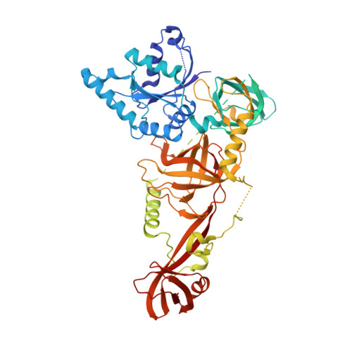
    Eukaryotic small ribosomal subunits are first assembled into 90S pre-ribosomes. The complete 90S is a gigantic complex with a molecular mass of approximately five megadaltons. Here, we report the nearly complete architecture of Saccharomyces cerevisiae 90S determined from three cryo-electron microscopy single particle reconstructions at 4.5 to 8.7 angstrom resolution. The majority of the density maps were modeled and assigned to specific RNA and protein components. The nascent ribosome is assembled into isolated native-like substructures that are stabilized by abundant assembly factors. The 5' external transcribed spacer and U3 snoRNA nucleate a large subcomplex that scaffolds the nascent ribosome. U3 binds four sites of pre-rRNA, including a novel site on helix 27 but not the 3' side of the central pseudoknot, and crucially organizes the 90S structure. The 90S model provides significant insight into the principle of small subunit assembly and the function of assembly factors.


  • Organizational Affiliation
    • PTN Joint Graduate Program, School of Life Sciences, Tsinghua University, Beijing, China.

Macromolecule Content 

  • Total Structure Weight: 82.88 kDa 
  • Atom Count: 4,397 
  • Modeled Residue Count: 531 
  • Deposited Residue Count: 709 
  • Unique protein chains: 1

Macromolecules

Find similar proteins by:|  3D Structure
Entity ID: 1
MoleculeChains  Sequence LengthOrganismDetailsImage
Ribosome biogenesis protein TSR1709Saccharomyces cerevisiae S288CMutation(s): 0 
Gene Names: TSR1YDL060W
UniProt
Find proteins for Q07381 (Saccharomyces cerevisiae (strain ATCC 204508 / S288c))
Explore Q07381 
Go to UniProtKB:  Q07381
Entity Groups
Sequence Clusters30% Identity50% Identity70% Identity90% Identity95% Identity100% Identity
UniProt GroupQ07381
Sequence Annotations
Expand
Reference Sequence

Small Molecules

Ligands 1 Unique
IDChains Name / Formula / InChI Key2D Diagram3D Interactions
SO4

Query on SO4


Download:Ideal Coordinates CCD File
B [auth A]
C [auth A]
D [auth A]
E [auth A]
F [auth A]
B [auth A],
C [auth A],
D [auth A],
E [auth A],
F [auth A],
G [auth A],
H [auth A],
I [auth A],
J [auth A],
K [auth A]
SULFATE ION
O4 S
QAOWNCQODCNURD-UHFFFAOYSA-L

Experimental Data & Validation

Experimental Data

  • Method: X-RAY DIFFRACTION
  • Resolution: 2.81 Å
  • R-Value Free:  0.274 (Depositor), 0.272 (DCC) 
  • R-Value Work:  0.214 (Depositor), 0.218 (DCC) 
  • R-Value Observed: 0.217 (Depositor) 
Space Group: P 21 21 21
Unit Cell:
Length ( Å )Angle ( ˚ )
a = 65.374α = 90
b = 116.344β = 90
c = 119.463γ = 90
Software Package:
Software NamePurpose
HKL-2000data scaling
PHENIXrefinement
PDB_EXTRACTdata extraction
HKL-2000data reduction
PHENIXphasing

Structure Validation

View Full Validation Report


Entry History 

Deposition Data

Revision History  (Full details and data files)

  • Version 1.0: 2017-06-28
    Type: Initial release
  • Version 1.1: 2024-03-20
    Changes: Data collection, Database references