5WWK | pdb_00005wwk

Highly stable green fluorescent protein


Experimental Data Snapshot

  • Method: X-RAY DIFFRACTION
  • Resolution: 3.20 Å
  • R-Value Free: 
    0.275 (Depositor), 0.274 (DCC) 
  • R-Value Work: 
    0.221 (Depositor), 0.221 (DCC) 
  • R-Value Observed: 
    0.226 (Depositor) 

Starting Model: experimental
View more details

wwPDB Validation 3D Report Full Report

Validation slider image for 5WWK

This is version 2.0 of the entry. See complete history

Literature

Excited State Electronic Interconversion and Structural Transformation of Engineered Red-Emitting Green Fluorescent Protein Mutant.

Augustine, G.Raghavan, S.NumbiRamudu, K.Easwaramoorthi, S.Shanmugam, G.Seetharani Murugaiyan, J.Gunasekaran, K.Govind, C.Karunakaran, V.Ayyadurai, N.

(2019) J Phys Chem B 123: 2316-2324

  • DOI: https://doi.org/10.1021/acs.jpcb.8b10516
  • Primary Citation Related Structures: 
    5WWK

  • PubMed Abstract: 

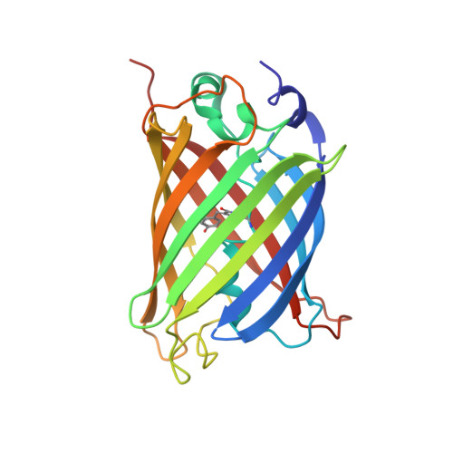
    Red fluorescent proteins with a large Stokes shift offer a limited autofluorescence background and are used in deep tissue imaging. Here, by introducing the free amino group in Aequorea victoria, the electrostatic charges of the p-hydroxybenzylidene imidazolinone chromophore of green fluorescent protein (GFP) have been altered resulting in an unusual, 85 nm red-shifted fluorescence. The structural and biophysical analysis suggested that the red shift is due to positional shift occupancy of Glu222 and Arg96, resulting in extended conjugation and a relaxed chromophore. Femtosecond transient absorption spectra exhibited that the excited state relaxation dynamics of red-shifted GFP (rGFP) (τ 4 = 234 ps) are faster compared to the A. victoria green fluorescent protein (τ 4 = 3.0 ns). The nanosecond time-resolved emission spectra of rGFP reveal the continuous spectral shift during emission by a solvent reorientation in the chromophore. Finally, the molecular dynamics simulations revealed the rearrangement of the hydrogen bond interactions in the chromophore vicinity, reshaping the symmetric distribution of van der Waals space to fine tune the GFP structure resulting from highly red-shifted rGFP.


  • Organizational Affiliation
    • Department of Biochemistry and Biotechnology , Council of Scientific and Industrial Research-Central Leather Research Institute (CSIR-CLRI) , Chennai 600 020 , India.

Macromolecule Content 

  • Total Structure Weight: 155.26 kDa 
  • Atom Count: 10,934 
  • Modeled Residue Count: 1,374 
  • Deposited Residue Count: 1,374 
  • Unique protein chains: 1

Macromolecules

Find similar proteins by:|  3D Structure
Entity ID: 1
MoleculeChains  Sequence LengthOrganismDetailsImage
Green fluorescent protein
A, B, C, D, E
A, B, C, D, E, F
229Aequorea victoriaMutation(s): 0 
UniProt
Find proteins for A0A059PIQ0 (Aequorea victoria)
Explore A0A059PIQ0 
Go to UniProtKB:  A0A059PIQ0
Entity Groups
Sequence Clusters30% Identity50% Identity70% Identity90% Identity95% Identity100% Identity
UniProt GroupA0A059PIQ0
Sequence Annotations
Expand
Reference Sequence

Small Molecules

Modified Residues  1 Unique
IDChains TypeFormula2D DiagramParent
CR2
Query on CR2
A, B, C, D, E
A, B, C, D, E, F
L-PEPTIDE LINKINGC13 H13 N3 O4GLY, TYR, GLY

Experimental Data & Validation

Experimental Data

  • Method: X-RAY DIFFRACTION
  • Resolution: 3.20 Å
  • R-Value Free:  0.275 (Depositor), 0.274 (DCC) 
  • R-Value Work:  0.221 (Depositor), 0.221 (DCC) 
  • R-Value Observed: 0.226 (Depositor) 
Space Group: P 21 21 21
Unit Cell:
Length ( Å )Angle ( ˚ )
a = 57.1α = 90
b = 95.976β = 90
c = 269.622γ = 90
Software Package:
Software NamePurpose
PHENIXrefinement
Aimlessdata scaling
PHASERphasing
PDB_EXTRACTdata extraction
XDSdata reduction
PHASERphasing

Structure Validation

View Full Validation Report


Entry History 

Deposition Data

Revision History  (Full details and data files)

  • Version 1.0: 2017-12-13
    Type: Initial release
  • Version 1.1: 2019-08-14
    Changes: Data collection, Database references
  • Version 1.2: 2019-11-06
    Changes: Data collection, Derived calculations
  • Version 1.3: 2023-11-22
    Changes: Data collection, Database references, Refinement description
  • Version 1.4: 2024-10-30
    Changes: Structure summary
  • Version 2.0: 2026-03-18
    Changes: Polymer sequence