5WW8 | pdb_00005ww8

Crystal structure of the second DNA-Binding protein under starvation from Mycobacterium smegmatis soaked with iron in the ratio of 480 iron atoms per dodecamer


Experimental Data Snapshot

  • Method: X-RAY DIFFRACTION
  • Resolution: 2.04 Å
  • R-Value Free: 
    0.210 (Depositor), 0.210 (DCC) 
  • R-Value Work: 
    0.177 (Depositor), 0.178 (DCC) 
  • R-Value Observed: 
    0.179 (Depositor) 

wwPDB Validation 3D Report Full Report

Validation slider image for 5WW8

This is version 1.1 of the entry. See complete history

Literature

Flexible aspartates propel iron to the ferroxidation sites along pathways stabilized by a conserved arginine in Dps proteins from Mycobacterium smegmatis

Williams, S.M.Chatterji, D.

(2017) Metallomics 9: 685-698

  • DOI: https://doi.org/10.1039/c7mt00008a
  • Primary Citation Related Structures: 
    5WW3, 5WW4, 5WW5, 5WW6, 5WW7, 5WW8, 5WW9

  • PubMed Abstract: 

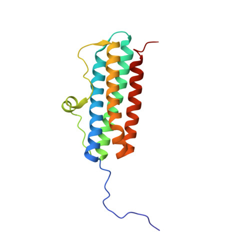
    DNA-binding proteins under starvation (Dps) are dodecameric nano-compartments for iron oxidation and storage in bacterial cells. These proteins have roughly spherical structures with a hollow interior where iron is stored. Through mutational analysis of a conserved arginine residue in the second Dps protein from Mycobacterium smegmatis, we have identified residues which stabilize the interfaces between the iron entry and ferroxidation sites. Also, we have used X-ray crystallography to determine the structures of co-crystals of iron and Dps in varying proportions and compare the changes in these ligand-bound forms with respect to the apo-protein. The iron-loaded proteins of low, medium and high iron-bound forms were found to exhibit aspartate residues with alternate conformations, some of which could be directly linked to the sites of ferroxidation and iron entry. We conclude that the increased flexibility of aspartates in the presence of iron facilitates its movement from the entry site to the ferroxidaton site, and the two active sites are stabilized by the interactions of a conserved arginine residue R73.


  • Organizational Affiliation
    • Molecular Biophysics Unit, Indian Institute of Science, Bangalore - 560 012, India. swilliams@mbu.iisc.ernet.in.

Macromolecule Content 

  • Total Structure Weight: 71.99 kDa 
  • Atom Count: 5,262 
  • Modeled Residue Count: 641 
  • Deposited Residue Count: 644 
  • Unique protein chains: 1

Macromolecules

Find similar proteins by:|  3D Structure
Entity ID: 1
MoleculeChains  Sequence LengthOrganismDetailsImage
Putative starvation-induced DNA protecting protein/Ferritin and Dps
A, B, C, D
161Mycolicibacterium smegmatis MC2 155Mutation(s): 0 
Gene Names: MSMEG_3242MSMEI_3159
UniProt
Find proteins for A0QXB7 (Mycolicibacterium smegmatis (strain ATCC 700084 / mc(2)155))
Explore A0QXB7 
Go to UniProtKB:  A0QXB7
Entity Groups
Sequence Clusters30% Identity50% Identity70% Identity90% Identity95% Identity100% Identity
UniProt GroupA0QXB7
Sequence Annotations
Expand
Reference Sequence

Small Molecules

Ligands 4 Unique
IDChains Name / Formula / InChI Key2D Diagram3D Interactions
FE2

Query on FE2


Download:Ideal Coordinates CCD File
F [auth A],
L [auth C],
O [auth D],
P [auth D]
FE (II) ION
Fe
CWYNVVGOOAEACU-UHFFFAOYSA-N
FE

Query on FE


Download:Ideal Coordinates CCD File
E [auth A],
I [auth B],
K [auth C],
N [auth D]
FE (III) ION
Fe
VTLYFUHAOXGGBS-UHFFFAOYSA-N
CL

Query on CL


Download:Ideal Coordinates CCD File
H [auth A],
M [auth C]
CHLORIDE ION
Cl
VEXZGXHMUGYJMC-UHFFFAOYSA-M
MG

Query on MG


Download:Ideal Coordinates CCD File
G [auth A],
J [auth B]
MAGNESIUM ION
Mg
JLVVSXFLKOJNIY-UHFFFAOYSA-N

Experimental Data & Validation

Experimental Data

  • Method: X-RAY DIFFRACTION
  • Resolution: 2.04 Å
  • R-Value Free:  0.210 (Depositor), 0.210 (DCC) 
  • R-Value Work:  0.177 (Depositor), 0.178 (DCC) 
  • R-Value Observed: 0.179 (Depositor) 
Space Group: H 3 2
Unit Cell:
Length ( Å )Angle ( ˚ )
a = 89.7α = 90
b = 89.7β = 90
c = 420.842γ = 120
Software Package:
Software NamePurpose
REFMACrefinement
MOSFLMdata reduction
SCALAdata scaling
REFMACphasing

Structure Validation

View Full Validation Report


Entry History 

Deposition Data

Revision History  (Full details and data files)

  • Version 1.0: 2017-08-09
    Type: Initial release
  • Version 1.1: 2024-03-20
    Changes: Data collection, Database references, Derived calculations