5WS2 | pdb_00005ws2

Crystal structure of mpy-RNase J (mutant S247A), an archaeal RNase J from Methanolobus psychrophilus R15, complex with RNA


Experimental Data Snapshot

  • Method: X-RAY DIFFRACTION
  • Resolution: 2.40 Å
  • R-Value Free: 
    0.208 (Depositor), 0.209 (DCC) 
  • R-Value Work: 
    0.173 (Depositor), 0.176 (DCC) 
  • R-Value Observed: 
    0.174 (Depositor) 

Starting Model: experimental
View more details

wwPDB Validation 3D Report Full Report

Validation slider image for 5WS2

This is version 1.1 of the entry. See complete history

Literature

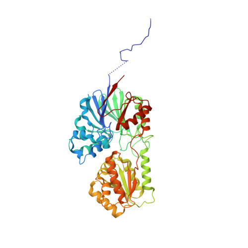
New molecular insights into an archaeal RNase J reveal a conserved processive exoribonucleolysis mechanism of the RNase J family

Zheng, X.Feng, N.Li, D.Dong, X.Li, J.

(2017) Mol Microbiol 106: 351-366

  • DOI: https://doi.org/10.1111/mmi.13769
  • Primary Citation Related Structures: 
    5WS2

  • PubMed Abstract: 

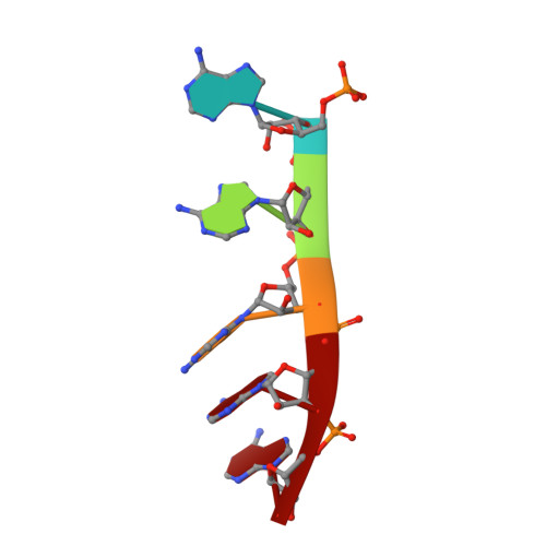
    RNase J, a prokaryotic 5'-3' exo/endoribonuclease, contributes to mRNA decay, rRNA maturation and post-transcriptional regulation. Yet the processive-exoribonucleolysis mechanism remains obscure. Here, we solved the first RNA-free and RNA-bound structures of an archaeal RNase J, and through intensive biochemical studies provided detailed mechanistic insights into the catalysis and processivity. Distinct dimerization/tetramerization patterns were observed for archaeal and bacterial RNase Js, and unique archaeal Loops I and II were found involved in RNA interaction. A hydrogen-bond-network was identified for the first time that assists catalysis by facilitating efficient proton transfer in the catalytic center. A conserved 5'-monophosphate-binding pocket that coordinates the RNA 5'-end ensures the 5'-monophosphate preferential exoribonucleolysis. To achieve exoribonucleolytic processivity, the 5'-monophosphate-binding pocket and nucleotide +4 binding site anchor RNA within the catalytic track; the 5'-capping residue Leu37 of the sandwich pocket coupled with the 5'-monophosphate-binding pocket are dedicated to translocating and controlling the RNA orientation for each exoribonucleolytic cycle. The processive-exoribonucleolysis mechanism was verified as conserved in bacterial RNase J and also exposes striking parallels with the non-homologous eukaryotic 5'-3' exoribonuclease, Xrn1. The findings in this work shed light on not only the molecular mechanism of the RNase J family, but also the evolutionary convergence of divergent exoribonucleases.


  • Organizational Affiliation
    • State Key Laboratory of Microbial Resources, Institute of Microbiology, Chinese Academy of Sciences, No.1 Beichen West Road, Chaoyang District, Beijing 100101, China.

Macromolecule Content 

  • Total Structure Weight: 108.66 kDa 
  • Atom Count: 7,790 
  • Modeled Residue Count: 936 
  • Deposited Residue Count: 950 
  • Unique protein chains: 1
  • Unique nucleic acid chains: 1

Macromolecules


Find similar proteins by:|  3D Structure
Entity ID: 1
MoleculeChains  Sequence LengthOrganismDetailsImage
Ribonuclease J
A, B
470Methanolobus psychrophilus R15Mutation(s): 1 
Gene Names: rnjMpsy_0886
EC: 3.1
Find similar nucleic acids by:  Sequence
Entity ID: 2
MoleculeChains LengthOrganismImage
RNA (5'-R(P*AP*AP*AP*AP*A)-3')
C, D
5unidentified
Sequence Annotations
Expand
Reference Sequence

Small Molecules

Ligands 2 Unique
IDChains Name / Formula / InChI Key2D Diagram3D Interactions
SO4

Query on SO4


Download:Ideal Coordinates CCD File
G [auth A]
H [auth A]
I [auth A]
L [auth B]
M [auth B]
G [auth A],
H [auth A],
I [auth A],
L [auth B],
M [auth B],
N [auth B],
O [auth B],
P [auth B],
Q [auth C]
SULFATE ION
O4 S
QAOWNCQODCNURD-UHFFFAOYSA-L
ZN

Query on ZN


Download:Ideal Coordinates CCD File
E [auth A],
F [auth A],
J [auth B],
K [auth B]
ZINC ION
Zn
PTFCDOFLOPIGGS-UHFFFAOYSA-N

Experimental Data & Validation

Experimental Data

  • Method: X-RAY DIFFRACTION
  • Resolution: 2.40 Å
  • R-Value Free:  0.208 (Depositor), 0.209 (DCC) 
  • R-Value Work:  0.173 (Depositor), 0.176 (DCC) 
  • R-Value Observed: 0.174 (Depositor) 
Space Group: P 65 2 2
Unit Cell:
Length ( Å )Angle ( ˚ )
a = 168.61α = 90
b = 168.61β = 90
c = 165.868γ = 120
Software Package:
Software NamePurpose
PHENIXrefinement
HKL-2000data reduction
HKL-2000data scaling
PHASERphasing

Structure Validation

View Full Validation Report


Entry History 

Deposition Data

Revision History  (Full details and data files)

  • Version 1.0: 2017-12-06
    Type: Initial release
  • Version 1.1: 2023-11-08
    Changes: Data collection, Database references, Derived calculations, Refinement description