5WR9 | pdb_00005wr9

Crystal structure of hen egg-white lysozyme


Experimental Data Snapshot

  • Method: X-RAY DIFFRACTION
  • Resolution: 1.80 Å
  • R-Value Free: 
    0.184 (Depositor) 
  • R-Value Work: 
    0.175 (Depositor) 
  • R-Value Observed: 
    0.175 (Depositor) 

wwPDB Validation 3D Report Full Report

Validation slider image for 5WR9

This is version 1.3 of the entry. See complete history

Literature

Hydroxyethyl cellulose matrix applied to serial crystallography

Sugahara, M.Nakane, T.Masuda, T.Suzuki, M.Inoue, S.Song, C.Tanaka, R.Nakatsu, T.Mizohata, E.Yumoto, F.Tono, K.Joti, Y.Kameshima, T.Hatsui, T.Yabashi, M.Nureki, O.Numata, K.Nango, E.Iwata, S.

(2017) Sci Rep 7: 703-703

  • DOI: https://doi.org/10.1038/s41598-017-00761-0
  • Primary Citation Related Structures: 
    5WR8, 5WR9, 5WRA, 5WRB, 5WRC

  • PubMed Abstract: 

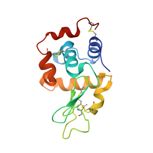
    Serial femtosecond crystallography (SFX) allows structures of proteins to be determined at room temperature with minimal radiation damage. A highly viscous matrix acts as a crystal carrier for serial sample loading at a low flow rate that enables the determination of the structure, while requiring consumption of less than 1 mg of the sample. However, a reliable and versatile carrier matrix for a wide variety of protein samples is still elusive. Here we introduce a hydroxyethyl cellulose-matrix carrier, to determine the structure of three proteins. The de novo structure determination of proteinase K from single-wavelength anomalous diffraction (SAD) by utilizing the anomalous signal of the praseodymium atom was demonstrated using 3,000 diffraction images.


  • Organizational Affiliation
    • RIKEN SPring-8 Center, 1-1-1 Kouto, Sayo-cho, Sayo-gun, Hyogo, 679-5148, Japan. msuga@spring8.or.jp.

Macromolecule Content 

  • Total Structure Weight: 14.39 kDa 
  • Atom Count: 1,078 
  • Modeled Residue Count: 129 
  • Deposited Residue Count: 129 
  • Unique protein chains: 1

Macromolecules

Find similar proteins by:|  3D Structure
Entity ID: 1
MoleculeChains  Sequence LengthOrganismDetailsImage
Lysozyme C129Gallus gallusMutation(s): 0 
Gene Names: LYZ
EC: 3.2.1.17
UniProt
Find proteins for P00698 (Gallus gallus)
Explore P00698 
Go to UniProtKB:  P00698
Entity Groups
Sequence Clusters30% Identity50% Identity70% Identity90% Identity95% Identity100% Identity
UniProt GroupP00698
Sequence Annotations
Expand
Reference Sequence

Experimental Data & Validation

Experimental Data

  • Method: X-RAY DIFFRACTION
  • Resolution: 1.80 Å
  • R-Value Free:  0.184 (Depositor) 
  • R-Value Work:  0.175 (Depositor) 
  • R-Value Observed: 0.175 (Depositor) 
Space Group: P 43 21 2
Unit Cell:
Length ( Å )Angle ( ˚ )
a = 80α = 90
b = 80β = 90
c = 38.4γ = 90
Software Package:
Software NamePurpose
PHENIXrefinement
CrystFELdata processing
CrystFELdata scaling
Cootmodel building

Structure Validation

View Full Validation Report


Entry History 

Deposition Data

Revision History  (Full details and data files)

  • Version 1.0: 2017-12-06
    Type: Initial release
  • Version 1.1: 2018-01-24
    Changes: Data collection
  • Version 1.2: 2023-09-06
    Changes: Data collection, Database references, Derived calculations
  • Version 1.3: 2024-10-16
    Changes: Structure summary