5WR4 | pdb_00005wr4

Thermolysin, SFX unliganded form with oil-based carrier


Experimental Data Snapshot

  • Method: X-RAY DIFFRACTION
  • Resolution: 2.10 Å
  • R-Value Free: 
    0.188 (Depositor), 0.188 (DCC) 
  • R-Value Work: 
    0.150 (Depositor), 0.155 (DCC) 
  • R-Value Observed: 
    0.152 (Depositor) 

Starting Model: experimental
View more details

wwPDB Validation 3D Report Full Report

Validation slider image for 5WR4

This is version 1.3 of the entry. See complete history

Literature

Protein-ligand complex structure from serial femtosecond crystallography using soaked thermolysin microcrystals and comparison with structures from synchrotron radiation

Naitow, H.Matsuura, Y.Tono, K.Joti, Y.Kameshima, T.Hatsui, T.Yabashi, M.Tanaka, R.Tanaka, T.Sugahara, M.Kobayashi, J.Nango, E.Iwata, S.Kunishima, N.

(2017) Acta Crystallogr D Struct Biol 73: 702-709

  • DOI: https://doi.org/10.1107/S2059798317008919
  • Primary Citation Related Structures: 
    5WR2, 5WR3, 5WR4, 5WR5, 5WR6

  • PubMed Abstract: 

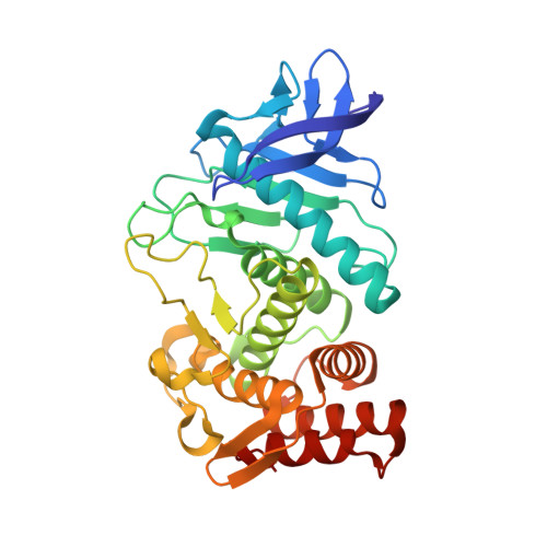
    Serial femtosecond crystallography (SFX) with an X-ray free-electron laser is used for the structural determination of proteins from a large number of microcrystals at room temperature. To examine the feasibility of pharmaceutical applications of SFX, a ligand-soaking experiment using thermolysin microcrystals has been performed using SFX. The results were compared with those from a conventional experiment with synchrotron radiation (SR) at 100 K. A protein-ligand complex structure was successfully obtained from an SFX experiment using microcrystals soaked with a small-molecule ligand; both oil-based and water-based crystal carriers gave essentially the same results. In a comparison of the SFX and SR structures, clear differences were observed in the unit-cell parameters, in the alternate conformation of side chains, in the degree of water coordination and in the ligand-binding mode.


  • Organizational Affiliation
    • Bio-Specimen Platform Group, RIKEN SPring-8 Center, Kouto, Sayo-cho, Sayo-gun, Hyogo 679-5148, Japan.

Macromolecule Content 

  • Total Structure Weight: 34.59 kDa 
  • Atom Count: 2,765 
  • Modeled Residue Count: 316 
  • Deposited Residue Count: 316 
  • Unique protein chains: 1

Macromolecules

Find similar proteins by:|  3D Structure
Entity ID: 1
MoleculeChains  Sequence LengthOrganismDetailsImage
Thermolysin316Geobacillus stearothermophilusMutation(s): 0 
Gene Names: nprSnprM
EC: 3.4.24.27
UniProt
Find proteins for P43133 (Geobacillus stearothermophilus)
Explore P43133 
Go to UniProtKB:  P43133
Entity Groups
Sequence Clusters30% Identity50% Identity70% Identity90% Identity95% Identity100% Identity
UniProt GroupP43133
Sequence Annotations
Expand
Reference Sequence

Experimental Data & Validation

Experimental Data

  • Method: X-RAY DIFFRACTION
  • Resolution: 2.10 Å
  • R-Value Free:  0.188 (Depositor), 0.188 (DCC) 
  • R-Value Work:  0.150 (Depositor), 0.155 (DCC) 
  • R-Value Observed: 0.152 (Depositor) 
Space Group: P 61 2 2
Unit Cell:
Length ( Å )Angle ( ˚ )
a = 93.67α = 90
b = 93.67β = 90
c = 130.95γ = 120
Software Package:
Software NamePurpose
PHENIXrefinement
CrystFELdata reduction
CrystFELdata scaling
PHENIXphasing

Structure Validation

View Full Validation Report


Entry History 

Deposition Data

Revision History  (Full details and data files)

  • Version 1.0: 2017-08-16
    Type: Initial release
  • Version 1.1: 2018-01-24
    Changes: Data collection
  • Version 1.2: 2023-09-06
    Changes: Data collection, Database references, Derived calculations
  • Version 1.3: 2023-11-08
    Changes: Refinement description