5WNX | pdb_00005wnx

DNA polymerase beta substrate complex with incoming 6-TdGTP


Experimental Data Snapshot

  • Method: X-RAY DIFFRACTION
  • Resolution: 2.55 Å
  • R-Value Free: 
    0.289 (Depositor), 0.287 (DCC) 
  • R-Value Work: 
    0.206 (Depositor), 0.206 (DCC) 
  • R-Value Observed: 
    0.214 (Depositor) 

Starting Model: experimental
View more details

wwPDB Validation 3D Report Full Report

Validation slider image for 5WNX

Ligand Structure Quality Assessment 


This is version 1.3 of the entry. See complete history

Literature

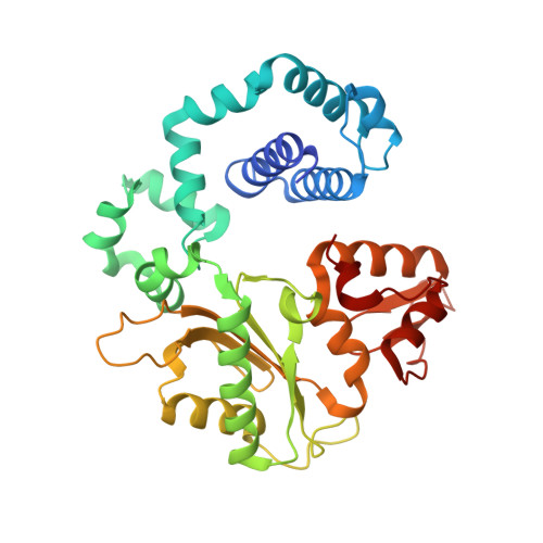
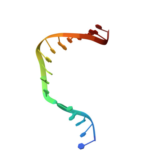
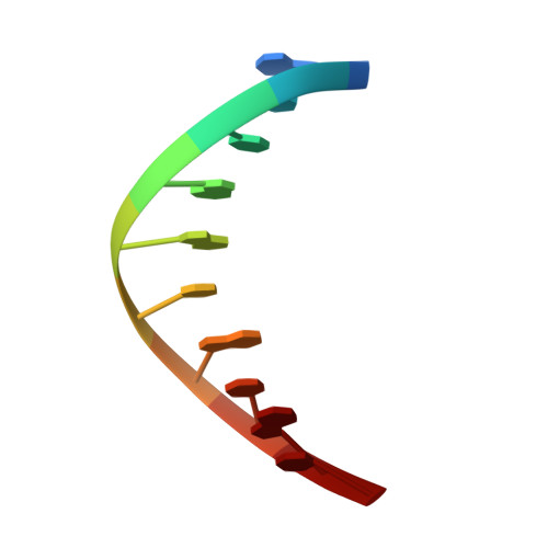
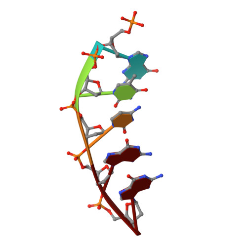
Structures of a DNA Polymerase Inserting Therapeutic Nucleotide Analogues.

Schaich, M.A.Smith, M.R.Cloud, A.S.Holloran, S.M.Freudenthal, B.D.

(2017) Chem Res Toxicol 30: 1993-2001

  • DOI: https://doi.org/10.1021/acs.chemrestox.7b00173
  • Primary Citation Related Structures: 
    5WNX, 5WNY, 5WNZ, 5WO0

  • PubMed Abstract: 

    Members of the nucleoside analogue class of cancer therapeutics compete with canonical nucleotides to disrupt numerous cellular processes, including nucleotide homeostasis, DNA and RNA synthesis, and nucleotide metabolism. Nucleoside analogues are triphosphorylated and subsequently inserted into genomic DNA, contributing to the efficacy of therapeutic nucleosides in multiple ways. In some cases, the altered base acts as a mutagen, altering the DNA sequence to promote cellular death; in others, insertion of the altered nucleotide triggers DNA repair pathways, which produce lethal levels of cytotoxic intermediates such as single and double stranded DNA breaks. As a prerequisite to many of these biological outcomes, the modified nucleotide must be accommodated in the DNA polymerase active site during nucleotide insertion. Currently, the molecular contacts that mediate DNA polymerase insertion of modified nucleotides remain unknown for multiple therapeutic compounds, despite decades of clinical use. To determine how modified bases are inserted into duplex DNA, we used mammalian DNA polymerase β (pol β) to visualize the structural conformations of four therapeutically relevant modified nucleotides, 6-thio-2'-deoxyguanosine-5'-triphosphate (6-TdGTP), 5-fluoro-2'-deoxyuridine-5'-triphosphate (5-FdUTP), 5-formyl-deoxycytosine-5'-triphosphate (5-FodCTP), and 5-formyl-deoxyuridine-5'-triphosphate (5-FodUTP). Together, the structures reveal a pattern in which the modified nucleotides utilize Watson-Crick base pairing interactions similar to that of unmodified nucleotides. The nucleotide modifications were consistently positioned in the major groove of duplex DNA, accommodated by an open cavity in pol β. These results provide novel information for the rational design of new therapeutic nucleoside analogues and a greater understanding of how modified nucleotides are tolerated by polymerases.


  • Organizational Affiliation
    • Department of Biochemistry and Molecular Biology, University of Kansas Medical Center , Kansas City, Kansas 66160, United States.

Macromolecule Content 

  • Total Structure Weight: 48.35 kDa 
  • Atom Count: 3,244 
  • Modeled Residue Count: 356 
  • Deposited Residue Count: 366 
  • Unique protein chains: 1
  • Unique nucleic acid chains: 3

Macromolecules


Find similar proteins by:|  3D Structure
Entity ID: 4
MoleculeChains  Sequence LengthOrganismDetailsImage
DNA polymerase betaD [auth A]335Homo sapiensMutation(s): 0 
Gene Names: POLB
EC: 2.7.7.7 (PDB Primary Data), 4.2.99 (PDB Primary Data), 4.2.99.18 (UniProt)
UniProt & NIH Common Fund Data Resources
Find proteins for P06746 (Homo sapiens)
Explore P06746 
Go to UniProtKB:  P06746
PHAROS:  P06746
GTEx:  ENSG00000070501 
Entity Groups
Sequence Clusters30% Identity50% Identity70% Identity90% Identity95% Identity100% Identity
UniProt GroupP06746
Sequence Annotations
Expand
Reference Sequence
Find similar nucleic acids by:  Sequence
Entity ID: 1
MoleculeChains LengthOrganismImage
DNA (5'-D(*CP*CP*GP*AP*CP*CP*GP*CP*GP*CP*AP*TP*CP*AP*GP*C)-3')A [auth T]16synthetic construct
Sequence Annotations
Expand
Reference Sequence
Find similar nucleic acids by:  Sequence
Entity ID: 2
MoleculeChains LengthOrganismImage
DNA (5'-D(*GP*CP*TP*GP*AP*TP*GP*CP*GP*C)-3')B [auth P]10synthetic construct
Sequence Annotations
Expand
Reference Sequence
Find similar nucleic acids by:  Sequence
Entity ID: 3
MoleculeChains LengthOrganismImage
DNA (5'-D(P*GP*TP*CP*GP*G)-3')C [auth D]5synthetic construct
Sequence Annotations
Expand
Reference Sequence

Small Molecules

Ligands 4 Unique
IDChains Name / Formula / InChI Key2D Diagram3D Interactions
SGT

Query on SGT


Download:Ideal Coordinates CCD File
H [auth A]2-amino-9-{2-deoxy-5-O-[(R)-hydroxy{[(R)-hydroxy(phosphonooxy)phosphoryl]oxy}phosphoryl]-beta-D-erythro-pentofuranosyl}-1,9-dihydro-6H-purine-6-thione
C10 H16 N5 O12 P3 S
BXZRFHUVVWIHMV-KVQBGUIXSA-N
CA

Query on CA


Download:Ideal Coordinates CCD File
I [auth A],
J [auth A]
CALCIUM ION
Ca
BHPQYMZQTOCNFJ-UHFFFAOYSA-N
CL

Query on CL


Download:Ideal Coordinates CCD File
G [auth A]CHLORIDE ION
Cl
VEXZGXHMUGYJMC-UHFFFAOYSA-M
NA

Query on NA


Download:Ideal Coordinates CCD File
E [auth A],
F [auth A]
SODIUM ION
Na
FKNQFGJONOIPTF-UHFFFAOYSA-N

Experimental Data & Validation

Experimental Data

  • Method: X-RAY DIFFRACTION
  • Resolution: 2.55 Å
  • R-Value Free:  0.289 (Depositor), 0.287 (DCC) 
  • R-Value Work:  0.206 (Depositor), 0.206 (DCC) 
  • R-Value Observed: 0.214 (Depositor) 
Space Group: P 1 21 1
Unit Cell:
Length ( Å )Angle ( ˚ )
a = 50.243α = 90
b = 79.203β = 107.36
c = 55.625γ = 90
Software Package:
Software NamePurpose
PHENIXrefinement
HKL-3000data reduction
HKL-3000data scaling
PHENIXphasing

Structure Validation

View Full Validation Report


Ligand Structure Quality Assessment 


Entry History 

& Funding Information

Deposition Data


Funding OrganizationLocationGrant Number
National Institutes of Health/National Institute of Environmental Health Sciences (NIH/NIEHS)United StatesES024432

Revision History  (Full details and data files)

  • Version 1.0: 2017-09-13
    Type: Initial release
  • Version 1.1: 2017-11-29
    Changes: Author supporting evidence, Database references
  • Version 1.2: 2019-12-18
    Changes: Author supporting evidence
  • Version 1.3: 2023-10-04
    Changes: Data collection, Database references, Derived calculations, Refinement description