5WM1 | pdb_00005wm1

Structure of the 10S (+)-trans-BP-dG modified Rev1 ternary complex


Experimental Data Snapshot

  • Method: X-RAY DIFFRACTION
  • Resolution: 1.85 Å
  • R-Value Free: 
    0.206 (Depositor), 0.213 (DCC) 
  • R-Value Work: 
    0.170 (Depositor), 0.179 (DCC) 
  • R-Value Observed: 
    0.172 (Depositor) 

Starting Model: experimental
View more details

wwPDB Validation 3D Report Full Report

Validation slider image for 5WM1

Ligand Structure Quality Assessment 


This is version 1.3 of the entry. See complete history

Literature

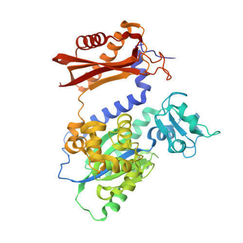
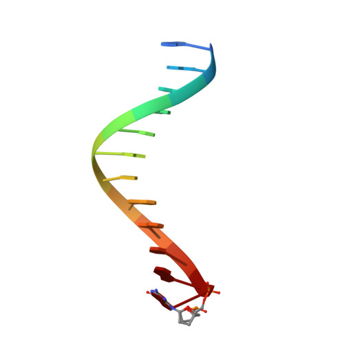
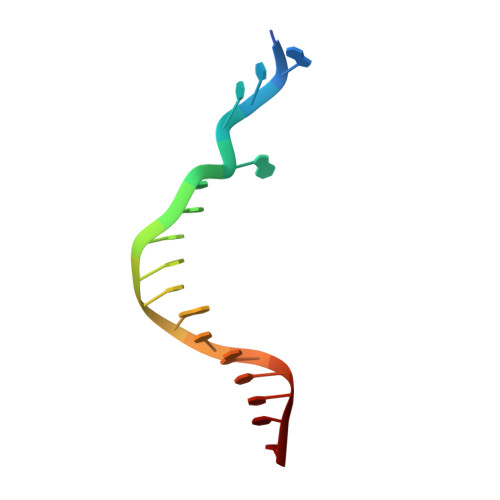
Mechanism of error-free replication across benzo[a]pyrene stereoisomers by Rev1 DNA polymerase.

Rechkoblit, O.Kolbanovskiy, A.Landes, H.Geacintov, N.E.Aggarwal, A.K.

(2017) Nat Commun 8: 965-965

  • DOI: https://doi.org/10.1038/s41467-017-01013-5
  • Primary Citation Related Structures: 
    5WM1, 5WM8, 5WMB

  • PubMed Abstract: 

    Benzo[a]pyrene (BP) is a carcinogen in cigarette smoke which, after metabolic activation, can react with the exocyclic N 2 amino group of guanine to generate four stereoisomeric BP-N 2 -dG adducts. Rev1 is unique among translesion synthesis DNA polymerases in employing a protein-template-directed mechanism of DNA synthesis opposite undamaged and damaged guanine. Here we report high-resolution structures of yeast Rev1 with three BP-N 2 -dG adducts, namely the 10S (+)-trans-BP-N 2 -dG, 10R (+)-cis-BP-N 2 -dG, and 10S ( - )-cis-BP-N 2 -dG. Surprisingly, in all three structures, the bulky and hydrophobic BP pyrenyl residue is entirely solvent-exposed in the major groove of the DNA. This is very different from the adduct alignments hitherto observed in free or protein-bound DNA. All complexes are well poised for dCTP insertion. Our structures provide a view of cis-BP-N 2 -dG adducts in a DNA polymerase active site, and offer a basis for understanding error-free replication of the BP-derived stereoisomeric guanine adducts.Benzo[a]pyrene (BP) is a carcinogen in cigarette smoke that upon metabolic activation reacts with guanine. Here, the authors present the structures of the translesion DNA synthesis polymerase Rev1 in complex with three of the four possible stereoisomeric BP-N 2 -dG adducts, which gives insights how Rev1 achieves error-free replication.


  • Organizational Affiliation
    • Department of Pharmacological Sciences, Icahn School of Medicine at Mount Sinai, Box 1677, 1425 Madison Avenue, New York, NY, 10029, USA. olga.rechkoblit@mssm.edu.

Macromolecule Content 

  • Total Structure Weight: 60.22 kDa 
  • Atom Count: 4,555 
  • Modeled Residue Count: 462 
  • Deposited Residue Count: 463 
  • Unique protein chains: 1
  • Unique nucleic acid chains: 2

Macromolecules


Find similar proteins by:|  3D Structure
Entity ID: 3
MoleculeChains  Sequence LengthOrganismDetailsImage
DNA repair protein REV1C [auth A]434Saccharomyces cerevisiae S288CMutation(s): 0 
Gene Names: REV1YOR346WO6339
EC: 2.7.7
UniProt
Find proteins for P12689 (Saccharomyces cerevisiae (strain ATCC 204508 / S288c))
Explore P12689 
Go to UniProtKB:  P12689
Entity Groups
Sequence Clusters30% Identity50% Identity70% Identity90% Identity95% Identity100% Identity
UniProt GroupP12689
Sequence Annotations
Expand
Reference Sequence
Find similar nucleic acids by:  Sequence
Entity ID: 1
MoleculeChains LengthOrganismImage
DNA (5'-D(*GP*GP*GP*GP*TP*GP*TP*GP*GP*TP*AP*(DDG))-3')A [auth P]12synthetic construct
Sequence Annotations
Expand
Reference Sequence
Find similar nucleic acids by:  Sequence
Entity ID: 2
MoleculeChains LengthOrganismImage
DNA (5'-D(P*AP*TP*CP*GP*CP*TP*AP*CP*CP*AP*CP*AP*CP*CP*CP*C)-3')B [auth T]17synthetic construct
Sequence Annotations
Expand
Reference Sequence

Small Molecules

Ligands 6 Unique
IDChains Name / Formula / InChI Key2D Diagram3D Interactions
DCP

Query on DCP


Download:Ideal Coordinates CCD File
J [auth A]2'-DEOXYCYTIDINE-5'-TRIPHOSPHATE
C9 H16 N3 O13 P3
RGWHQCVHVJXOKC-SHYZEUOFSA-N
BAP

Query on BAP


Download:Ideal Coordinates CCD File
E [auth T]1,2,3-TRIHYDROXY-1,2,3,4-TETRAHYDROBENZO[A]PYRENE
C20 H16 O3
GFANZDFKCCJYRF-NSISKUIASA-N
PEG

Query on PEG


Download:Ideal Coordinates CCD File
W [auth A]DI(HYDROXYETHYL)ETHER
C4 H10 O3
MTHSVFCYNBDYFN-UHFFFAOYSA-N
GOL

Query on GOL


Download:Ideal Coordinates CCD File
K [auth A],
L [auth A],
M [auth A],
N [auth A],
O [auth A]
GLYCEROL
C3 H8 O3
PEDCQBHIVMGVHV-UHFFFAOYSA-N
EDO

Query on EDO


Download:Ideal Coordinates CCD File
F [auth T]
P [auth A]
Q [auth A]
R [auth A]
S [auth A]
F [auth T],
P [auth A],
Q [auth A],
R [auth A],
S [auth A],
T [auth A],
U [auth A],
V [auth A]
1,2-ETHANEDIOL
C2 H6 O2
LYCAIKOWRPUZTN-UHFFFAOYSA-N
MG

Query on MG


Download:Ideal Coordinates CCD File
D [auth P],
G [auth A],
H [auth A],
I [auth A]
MAGNESIUM ION
Mg
JLVVSXFLKOJNIY-UHFFFAOYSA-N

Experimental Data & Validation

Experimental Data

  • Method: X-RAY DIFFRACTION
  • Resolution: 1.85 Å
  • R-Value Free:  0.206 (Depositor), 0.213 (DCC) 
  • R-Value Work:  0.170 (Depositor), 0.179 (DCC) 
  • R-Value Observed: 0.172 (Depositor) 
Space Group: P 21 21 21
Unit Cell:
Length ( Å )Angle ( ˚ )
a = 64.002α = 90
b = 64.879β = 90
c = 130.652γ = 90
Software Package:
Software NamePurpose
REFMACrefinement
HKL-2000data reduction
HKL-2000data scaling
PHENIXphasing

Structure Validation

View Full Validation Report


Ligand Structure Quality Assessment 


Entry History 

& Funding Information

Deposition Data


Funding OrganizationLocationGrant Number
National Institutes of Health/National Institute of Environmental Health Sciences (NIH/NIEHS)United StatesR01 CA200575
National Institutes of Health/National Institute of Environmental Health Sciences (NIH/NIEHS)United StatesR01 ES024050

Revision History  (Full details and data files)

  • Version 1.0: 2017-10-25
    Type: Initial release
  • Version 1.1: 2017-11-01
    Changes: Database references
  • Version 1.2: 2019-12-18
    Changes: Author supporting evidence
  • Version 1.3: 2023-10-04
    Changes: Data collection, Database references, Derived calculations, Refinement description